
Page 215 - 2021_06-Haematologica-web
P. 215
Letters to the Editor
AB
CD
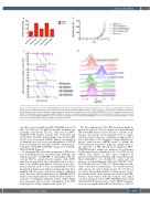
Figure 1. In vitro mutational screen of EBF1-PDGFRB reveals kinase domain mutations causing varying degrees of resistance to imatinib and dasatinib. (A) The proportion of T681I and N666S kinase domain mutations identified in EBF1-PDGFRB in vitro screens to different concentrations of imatinib and dasatinib. (B) Proliferation assays demonstrating the cytokine-independent proliferation of wild-type and mutant EBF1-PDGFRB Ba/F3 cells. (C) Drug-sensitivity profiles of Ba/F3 cells harboring wild-type and mutant EBF1-PDGFRB in response to imatinib, dasatinib and ponatinib. (D) Phosphorylation of STAT5 is elevated at basal in Ba/F3 cells harboring EBF1-PDGFRB and can be inhibited in wild-type but not mutant EBF1-PDGFRB in response to dasatinib. E-P: EBF1-PDGFRB; EV: empty vector; IL3: interleukin-3: WT: wild-type; pSTAT5: phosphorylated STAT5.
tion (IC50) values for wild-type EBF1-PDGFRB were 15.74 nM, 5.26 nM and 5.73 nM for imatinib, dasatinib and ponatinib, respectively. The IC50 values for the EBF1- PDGFRB T681I mutant isoform were 602.5 nM and 23.93 nM for imatinib and ponatinib, respectively, while the IC50 was not reached with the highest concentration of dasatinib used. Moreover, phosphorylation of STAT5 was not abrogated by dasatinib in Ba/F3 constructs har- boring the T681I EBF1-PDGFRB compared to wild-type EBF1-PDGFRB (Figure 1).
To understand the molecular mechanism of TKI resist- ance from KD mutations, we modeled the wild-type and mutant structures of PDGFRB in relationship with the relevant TKI. Co-crystal structure analysis of the T681I mutation demonstrated that substitution from a threo- nine to the bulkier hydrophobic isoleucine at the gate- keeper position leads to steric incompatibility between the ligand and the pocket, thus preventing dasatinib from binding both the active and inactive kinase conforma- tions. As for the N666S substitution, the PDGFRB N666S model demonstrated that the mutation likely disrupts a network of stabilizing hydrogen bonds, which might have long-range effects on the conformation of the ATP binding pocket (Online Supplementary Figure S1).
We then hypothesized that KD mutations might be present at very low levels at diagnosis in patients with EBF1-PDGFRB when assessed by more sensitive tech- nologies and emerge as the dominant clone at relapse under the selective pressure of therapy, as suggested by a few adult studies.8,9 We designed a droplet digital poly- merase chain reaction (ddPCR) assay to identify the T681I mutation in patients’ diagnostic samples prior to any exposure to a TKI. Among the 23 diagnostic EBF1- PDGFRB patients’ samples we analyzed, the gatekeeper T681I mutation was identified in 13% (n=3/23) by our ddPCR assay (Figure 2). This cohort comprised 13 patients enrolled on the Children’s Oncology Group ALL trials (AALL0232: n=1, AALL1131: n=12) and ten patients on United Kingdom ALL trials (UK ALL 97/99: n=3, UK ALL 2003: n=7) (Table 1). The median age of the entire cohort was 12 years (range, 8-16), and the median white blood cell count at diagnosis was 39.0 (17-80.7) x 109 cells/L. The median duration of follow-up was 60 (14- 81) months. None of the patients was treated with TKI. Baseline characteristics, leukemia response and clinical outcomes among the three EBF1-PDGFRB patients with subclonal T681I mutation detected by ddPCR at diagno- sis were not significantly different from those of the 20
haematologica | 2021; 106(8)
2243
