
Page 191 - 2021_06-Haematologica-web
P. 191
MDS progression involves cohesin and RAS mutations
stage, 210 mutations were detected, 159 of which were already known to be present in the MDS stage, at clonal or subclonal levels (128 were retained and 31 evolved during disease progression), while 51 were detected only at the second sampling. The most recurrently mutated genes at the sAML stage were similar to those noted at the MDS stage: TET2 (17 of 42, 40%), SF3B1 (13 of 42, 31%), TP53 (12 of 42, 29%), SRSF2 (ten of 42, 24%), DNMT3A (ten of 42, 24%), ASXL1 (nine of 42, 21%) and STAG2 (eight of 42, 19%). However, the NRAS gene (9 of 42, 21%) also stood out at this stage. It should be noted that 32 of 54 genes (59%) were mutated only in fewer than three (<7%) patients, highlighting the great heterogeneity in the mecha- nisms of disease evolution.
Regardless of World Health Organization diagnosis subtypes, patients progressing to secondary acute myeloid leukemia present a higher number of mutations than those that do not progress
In order to analyze the changes in clonal size and distri- bution during evolution to sAML, we compared the num- ber of mutations identified at diagnosis and at the second sampling in the discovery and control cohorts. The control cohort presented a median of three mutations at both sam- pling times (p10-p90: 2-4 in both), indicating no significant differences (P=0.449). By contrast, the discovery cohort had a median of four (p10-p90: 1-6) and five (p10-p90: 2-9) mutations at the first and second samplings, respectively, representing a highly significant increase in the number of mutations during disease progression (P<0.0001). Remarkably, the control and discovery cohorts had a similar number of mutations at the time of diagnosis (P=0.097), although a slight trend was observed, while patients who progressed showed a significantly higher number of muta- tions at the time of sAML than the control patients at the second sampling (P=0.027) (Figure 1A). Considering the dis- covery cohort patients by World Health Organization diag- nosis subtype (LR-MDS and HR-MDS) did not reveal any
significant differences in the number of mutations in patients progressing to sAML (P=0.588) (Figure 1B).
In order to further study what characterizes disease evo- lution, we compared the VAF of mutations at both times. Patients who evolved to sAML presented a significantly higher VAF median at second sampling (29.11% vs. 36.76%, P<0.0001) (Figure 1C). However, no differences were identified in the median VAF between each subtype at the time of diagnosis (P=0.528). (Figure 1D).
Therefore, taking all these results together, MDS patients, irrespective of their diagnostic subtype, displayed a greater genomic instability during disease progression than patients who did not evolve to sAML.
Mutational dynamics during the progression to secondary acute myeloid leukemia: clonal evolution
In order to study the mutational dynamics and identify which mutations could be involved in clonal evolution and play an important role during disease progression, the VAF of mutations detected at both times (follow- up/sAML vs. diagnosis) were compared in all patients of the discovery and control cohorts.
Four types of clonal dynamics were identified: type 1, in which mutations were initially present in the MDS stage, but whose VAF increased significantly in the sAML stage; type 2, mutations whose VAF significantly decreased; type 3, mutations that were newly acquired at the sAML stage; type 4, mutations that persisted with a similar allelic burden at both stages.
Stable mutations (Figure 2A, type 4, depicted in yellow) were detected in genes involved in the spliceosome and DNA methylation pathways, such as the splicing factor SRSF2 (diagnosis vs. sAML median VAF, P=0.4922) and the DNA methylation gene DNMT3A (diagnosis vs. sAML median VAF, P=0.7695) (Online Supplementary Figure S3).
Only a minority of the mutations detected at diagnosis showed a decrease in their allelic burden (Figure 2A, type
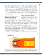
Figure 3. Model of clonal evolution during myelodysplastic syndrome progression to secondary acute myeloid leukemia using patient #43 as an example and apply- ing the Fishplot R package.29 In this patient diagnosed as RAEB-1, an myelodysplastic syndrome (MDS) founder clone was present at the time of diagnosis with typical myeloid mutations such as TET2 and SRSF2. This clone also harbored a mutation in STAG2, thus it triggered the acquisition of a subsequent mutation in a Ras pathway gene, namely NRAS. This clone expanded, driving the evolution of the disease.
haematologica | 2021; 106(8)
2219
