
Page 171 - 2021_06-Haematologica-web
P. 171
MYB bi-allelic targeting
ABC
DE
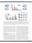
Figure 6. Vascular endothelial growth factor is a key cytokine for the early human hematopoiesis. (A) Human embryonic stem cells (hESC)-derived hematopoiesis on day 6 of differentiation is severely inhibited upon removal of vascular endothelial growth factor (VEGF) starting from day 3. Representative data of three experi- ments are shown. (B) Effect of the VEGF removal on the emergence of hematopoietic clonogenic progenitors during differentiation of H1 hESC (day 6 – day 16). Data are mean ± standard deviation (SD), n=4. (C) hESC-derived myeloid cells emerge normally on day 16 of differentiation in the absence of exogenous VEGF. Representative data of four experiments are shown. (D) Dynamics of hematopoietic marker expression during H1 hESC differentiation in the presence (open bars) or the absence of VEGF (colored bars). Data are presented as mean ± SD, n=3. (E) Day 20 phenotype of the MYB knockout cell lines differentiated in the absence of VEGF. Data are representative of three independent differentiation experiments.
independent early blood cells since these early cells are strongly suppressed by the VEGF removal (Figure 6A and D). Progenitor-independent monocyte/macrophages, granulocytes, and their immature precursors are likely to arise through the conversion of mesodermal or endothelial cells in a process similar to the aforementioned in vitro EHT. MYB inactivation severely affected the progenitor- independent myeloid cells with complete loss of granulo- cytes on day 20 of the VEGF-deprived differentiation (Figure 6E). Taken together, these results demonstrate that VEGF serves as a major mitogenic factor for the nascent human blood cells.
MYB is required for the development and proliferation of primitive progenitors
The progenitor defects upon MYB inactivation might be either due to the failure of progenitor proliferation in the methylcellulose assay or the interruption of progenitor development during the primary differentiation of hESC. In order to address this issue, we performed a series of res- cue experiments in MYB-null cells using a Tet-On expres- sion system PB-iDox that included a PiggyBac transposon vector25 bearing MYB cDNA under the TRE-CMV promot- er. One type of the doxycycline (DOX)-inducible vectors contained mCherry gene reporter attached to MYB cDNA
via an IRES element (Figure 7A and B). In CFU-C colonies that were generated from DOX-activated DKO cells trans- fected by the PB-MYB-mCherry construct, the red reporter was downregulated at the colony fringes containing more mature cells (Figure 7C). The transgene’s downregulation was even more profound than the silencing of MYB-Venus expression in the fringes (Online Supplementary Figure S7A). This observation suggests that the transgene’s expression is controlled by post-transcriptional regulatory mecha- nisms, possibly via microRNA,26 and the overexpression of MYB, a typical proto-oncogene, does not cause an uncontrolled proliferation of hematopoietic cells and pro- genitors. A time-course analysis of the PB-MYB transgene expression in DOX-treated DKO cells demonstrated stage-specific modulation of MYB mRNA levels further suggesting the post-transcriptional regulation by microRNA (Online Supplementary Figure S7B).
We have been able to rescue day 6 and day 12 clono- genic progenitors only when the transgenic MYB was induced by DOX both in the primary differentiation of hESC and the secondary methylcellulose cultures (Figure 7D; Online Supplementary Figure S7C). The most efficient was the recovery of BFU-EP on day 6 (Figure 7D) and myeloid progenitors on day 12 (Online Supplementary Figure S7C) whereas the rescue of CFU-mix progenitors
haematologica | 2021; 106(8)
2199
