
Page 158 - 2021_06-Haematologica-web
P. 158
G. Wu and C.C. Zhang
CAR stimulates Notch signaling
We sought to identify the underlying mechanism by which CAR mediates hematopoietic regeneration after stress. CAR binds to LNX, an E3 ligand of Numb,21 sug- gesting the function of CAR may be related to Numb. After 5-FU treatment, Numb protein levels in HSC grad- ually decreased, and the population with no Numb stain- ing increased from 5.7±0.3% on day 0 to 62.4±1.4% on day 5 (Figure 6A). On day 7, the Numb-negative popula- tion decreased to 6.8±1.5% (Figure 6A) as HSC had returned to homeostasis. Concordantly, the Numb mRNA levels decreased by more than 16 times on day 5 relative to pre-treatment levels and had returned to pre- treatment levels on day 7 (Figure 6A). HSC in WT mice entered the cell cycle earlier than did CAR cKO counter- parts after 5-FU treatment (Figure 5). At 1.5 days after 5- FU treatment, there were fewer LSKFC cells that did not stain for Numb in CAR cKO mice than WT mice (Figure 6B), whereas there were no differences detected before or 5 days later after 5-FU treatment (Online Supplementary Figure 10A). The Numb mRNA levels in CAR cKO mice were similar to those in WT mice at 1.5 days after 5-FU treatment (Online Supplementary Figure 10B), suggesting that CAR mediates the degradation of Numb protein but does not affect Numb mRNA. In addition, there was fewer Numb protein in CAR+ LSKFC cells than in CAR- LSKFC cells (Figure 6C). Because Numb specifically inhibits Notch signaling,26-28 these results suggest that Notch signaling is involved in CAR-mediated hematopoietic regeneration.
Notch signaling was previously reported to play an important role in hematopoietic regeneration.6,29,30 In
order to test how Notch signaling influences the function of CAR during regeneration, we treated mice with DAPT, an inhibitor of Notch signaling, with or without 300 mg/kg 5-FU. Whereas 5-FU treatment decreased sur- vival of CAR cKO mice relative to WT counterparts, there was no difference in survival between 5-FU-treated WT and CAR cKO mice when Notch signaling was blocked by DAPT (Figure 6D). This implies that CAR stimulates regeneration via Notch signaling. At 1.5 days after 5-FU treatment, 4.2% of LSKFC cells were Notch1 positive in CAR cKO mice compared to 8.7% in WT mice, whereas 27.7% and 27% of LSKFC cells in CAR cKO and WT mice were positive for Notch2, respective- ly (Figure 6E). This indicate that Notch1, but not Notch2, is involved in the function of CAR. There were signifi- cantly lower levels of hes1, hey1, and myc, all Notch1 tar- get genes, in CAR cKO mice compared to WT mice at 1.5 days after 5-FU treatment; there were no differences in levels of these mRNA prior to 5-FU treatment (Figure 6F). In addition, CAR cKO resulted in a defect in T-cell repop- ulation (Online Supplementary Figure 11), which further suggests a connection between CAR and Notch1, as Notch1 affects T-cell development.31 Notch1 target genes indeed increased right after 5-FU treatment (Online Supplementary Figure 12). With 5-FU treatment, CAR+ HSC had more surface Notch1 protein than CAR- HSC (Figure 6G). Overexpression of CAR in LSKFC cells in vitro also increased Notch1 target gene expression (Online Supplementary Figure 13). In order to further confirm that CAR stimulates Notch signaling, we overexpressed the dominant negative MAML (dnMAML)32-34 in 5-FU treated Lin- BM cells from UBC-Cre-ERT2/CARloxp/loxp mice
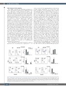
Figure 5. CAR stimulates hematopoietic stem cells proliferation after 5-fluorouracil treatment. Before and after 5-fluorouracil (5-FU) (150 mg/kg) treatment, cell cycle states of LSKFC cells were determined. The flow cytometry images are the representative results of bone marrow (BM) samples from wild-type (WT) or CAR conditional knockout (cKO) mice different times after 5-FU treatment, and the summaries of each time point after 5-FU treatment are on the right of flow cytometry images. The percentages of cell cycle states in total hematopoietic stem cells (HSC) (LSKFC) are indicated. Every groups include n=4-9 mice. G0: cell in G0 phase; G1; SG2M: cell in S, G2 and M phase; AP: cells in apoptosis. *P<0.05, **P<0.001.
2186
haematologica | 2021; 106(8)
