
Page 240 - 2021_07-Haematologica-web
P. 240
Letters to the Editor
from Becton Dickinson, BD; USA), CytoFlex and Navios (both from Beckman Coulter, BC; USA) flow cytometers. EuroFlow guidelines for machine performance monitor- ing were used.8 The immunophenotype of the tumor cells was analyzed with focus on markers applicable for MFC- MRD investigation.4,9 Online Supplementary Table S2 pro- vides a list of monoclonal antibodies used for MFC-MRD monitoring. CD22 and CD24 were additionally studied, mainly after the blinatumomab courses.10 Expression of surface antigens was deemed positive if the antigen was expressed on more than 20% of tumor cells.6 An increase or decrease of expression of each single antigen was defined as a change of the percentage of positive cells by more than 25%. Proportions of cases with stable and changed expression of each single antigen between CD19-negative and CD19-positive relapses were com- pared using the Fisher exact test.
The treatment outcomes of the studied patients are summarized in Figure 1. Thirty-nine patients achieved complete MFC-MRD-negative remission and three achieved bone marrow MFC-MRD-negativity, but with progression of extramedullary disease. These patients never had detectable leukemia in the bone marrow dur- ing follow-up, so they were excluded from analysis of immunophenotypic changes. Overall, modulation of antigen expression was studied in 48 patients with tumor blasts detectable in bone marrow at least once after a course of treatment with blinatumomab.
We focused separately on the status of CD19 expres- sion on leukemic cells (Figure 1), since this is the sole pos- sible immunophenotypic change directly linked to the administration of blinatumomab. Thirty patients experi- enced relapse (>5% of blasts cells by MFC). In 21 cases, leukemic cells at relapse were CD19-positive and in six
A
BC
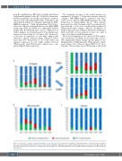
Figure 2. Frequency of changes in immunophenotype of leukemic blasts. (A-C) Frequency of changes in immunophenotype of leukemic blasts in relapsed cases (A), cases with detectable blasts in bone marrow only at a minimal residual disease level (B), and in resistant cases (C). Cases of “lineage switch” are not shown. *Differences in frequencies of antigen expression changes are statistically significant (P<0.05). MRD: minimal residual disease; MFC: multicolor flow cytometry.
2010
haematologica | 2021; 106(7)
