
Page 236 - 2021_07-Haematologica-web
P. 236
Letters to the Editor
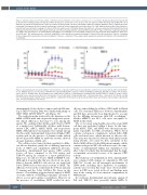
Figure 1. Activation-induced cell death activity of CD38 monclonal antibodies, in the absence and presence of crosslinker. Analyzed at 24 hoursa in Daudi cells by (A) annexin V staining and (B) at 5 daysb by CellTiter Glo Assay; no activity was seen in (C) LP-1 cells at 5 daysb by CellTiter Glo Assay and (D) MOLP-8 cells at 48 hoursa by annexin V staining and at 5 daysb by CellTiter Glo Assay. Daudi cells were plated with or without crosslinker (AffiniPure F(ab')2 fragment goat anti- human IgG, Fcγ fragment specific, in which endotoxin was removed; Jackson ImmunoResearch, West Grove, PA, USA). Antibody dilutions were added and incu- bated for 24 hours (annexin V staining) or 5 days (CellTiter Glo Luminescent Cell Viability Assay). After 24 hours, cells were washed and stained with Live/Dead (Invitrogen) followed by annexin V (BioLegend, San Diego, CA, USA) and analyzed by flow cytometry (FACSCanto). After 5 days, CellTiter Glo reagent was prepared according to the manufacturer’s recommendations and added to a second plate to assess viability. Luminescence was measured and normalized to “plate max” and “plate min”. This analysis was also conducted using MOLP-8 cells, with 48-hour (annexin V staining) or 5-day (CellTiter Glo) incubation. aData are a summary of three independent experiments performed in duplicate; bdata are a summary of three independent experiments performed in triplicate. ISA: isatuximab; XL: crosslinking.
Figure 2. Daratumumab demonstrated higher cytotoxicity than comparator CD38 monoclonal antibodies in whole blood assays with (A) LP-1 and (B) MOLP- 8 cells. Multiple myeloma cell lines LP-1 or MOLP-8 were labeled with DELFIA® europium solution (PerkinElmer, Pittsburgh, PA, USA) according to the manufac- turer’s protocol. Healthy donor blood samples were added with a titration of daratumumab, isatuximab (ISA) analog, TAK-079 analog, or isotype control. Europium release was measured after 3-hour incubation. Percent cytotoxicity = [(experimental lysis – min lysis)/(max lysis – min lysis)] x 100. Daudi cells did not effectively label with europium in this assay, which is a phenomenon that has been previously described. Data are shown as representative experiments (LP-1, n=6 donors; MOLP-8, n=5 donors).
AB
daratumumab. It also did not compete with the ISA ana- log or TAK-079 analog. Data were similar with gating on CD19–CD20–CD138+CD27dim cells (Figure 3C)
The results from this study add to the literature on the MOA of CD38 mAb and can provide insight into poten- tial clinical differences that may be seen among the agents. We confirmed that all three mAb bind to CD38 at a similar level. Additionally, all three mAb demon- strated CDC, ADCC, ADCP, AICD, and trogocytosis MOA. Although most mechanisms were similar among the three mAb, daratumumab demonstrated higher CDC activity and, in the presence of human serum (which allows all possible MOA for antibody activity), showed stronger depletion of MM cells.
The cell lines tested varied in their sensitivity to differ- ent effector functions, partly due to differing expression levels of CD38 and complement inhibitory proteins. Regardless, daratumumab had greater CDC activity across cell lines compared to the ISA analog and TAK-079 analog. In contrast to a report published by Jiang et al.,9 we did not observe AICD in MOLP-8 cells. Although it is unclear why this was observed, one possi- bility is that the MOLP-8 cells used in our study had lower CD38 levels or were more resistant to AICD. The difference between the results from the 24-hour annexin V staining and the 5-day CellTiter Glo Assay in the absence of crosslinking suggests that the impact of AICD over time without crosslinking is minimal; cells may be able to recover and continue to proliferate. However, in the presence of crosslinking, AICD resulted in more
effective tumor killing by all three CD38 mAb in Daudi cells. The structural differences between daratumumab and ISA have previously been hypothesized to account for the differing interactions with FcR crosslinking.15 Neither MOLP-8 nor LP-1 cells were susceptible to AICD in this study.
The ex vivo assay using healthy donor blood and MM cell lines was performed within 3 hours. At this time- point, ADCC, CDC, and ADCP were the major mecha- nisms responsible for MM cell ablation. Whole blood contains endogenous complement, natural killer (NK) cells, and monocytes, which function as effector cells. This assay was repeated at 24 hours with similar results using absolute cell counts by flow cytometry of the labeled MM cell lines. In the MM patient samples, which contain endogenous NK cells and monocytes, the supe- riority of daratumumab killing was maintained even after 3 days. It is likely that daratumumab had the high- est maximal cytotoxicity because of its superior CDC activity.
Our study has several limitations. First, daratumumab was compared with analogs of comparators, ISA and TAK-709. Second, not all anti-tumor mechanisms of CD38 mAb, including direct inhibition of enzymatic activity, were tested in this study.5 Last, observed differ- ences in CDC were tested in blood, and findings may vary in the setting of a bone marrow compartment in MM patients.
In conclusion, daratumumab and surrogate analogs of ISA and TAK-079 have generally similar MOA. It
2006
haematologica | 2021; 106(7)
