
Page 164 - 2021_07-Haematologica-web
P. 164
A.S. Zayac et al.
involvement. PFS was assessed locally as the time from diagno- sis until disease progression, recurrence, or death.18 Overall sur- vival (OS) was calculated from diagnosis until death or last fol- low-up.
Statistical analysis
We compared clinicopathological characteristics between groups using Fisher exact tests and evaluated factors associated with baseline CNS involvement by univariate and multivariable logistic regression (reporting adjusted odds ratios, aOR). Associations with survival were examined in proportional haz- ard models, first univariate, and then stratified by general BL risk factors identified in the same dataset, reporting hazard ratios (HR).16 The cumulative incidence of CNS recurrence was studied in competing-risk models that accounted for other events such as systemic recurrence or death from any cause, reporting sub distribution hazard ratios (SHR).19 To address missing data on PS (7%), stage (2%), HIV positivity (2%), LDH (7%), hemoglobin (5%), and albumin values (9%), we averaged model coefficients and standard errors from 15 datasets using multiple imputation by chained equations.20 The imputation model included all covariates and outcomes. Estimates report 95% confidence intervals (in square brackets), and two-sided P values <0.05 were considered statistically significant.
Results
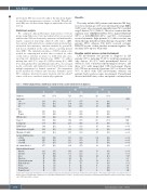
The study included 641 patients with untreated BL diag- nosed at a median age of 47 years (interquartile range [IQR], 34-59 years), who were predominantly male (76%) and had stage 4 disease (73%) (Table 1). The most common first-line regimens were CODOX-M/IVAC (30%), hyperCVAD/MA (30%), or DA-EPOCH-R (28%), and 90% of all patients received rituximab. Eight patients (1%) did not receive any chemotherapy. Intrathecal chemotherapy was given to 545 patients (85%) whereas 396 (62%) received systemic HDMTX as part of their first-line treatment regimen. The median follow-up was 45 months.
Baseline central nervous system involvement
CNS involvement was present at diagnosis in 120 patients (19%), including 97 (15%) with leptomeningeal- only disease, 20 (3%) with parenchymal disease (of whom 11 had concurrent leptomeningeal disease), and three (1%) with unspecified CNS involvement (Figure 1A). CSF was positive in 91 patients (14% of all cases, and 76% of those with CNS involvement), whereas ten patients had cavernous sinus involvement. Parenchymal disease included brain, ocular, and spinal cord invasion in
Table 1. Patient characteristics stratified by central nervous system involvement at diagnosis.
Numbers
All Baseline CNS involvement
No Yes
N (%) N (%) N (%)
641 (100) 521 (100) 120 (100)
P
0.747
0.37
<0.001 N/A 0.02 <0.001 <0.001 <0.001
<0.001 <0.001 <0.001
<0.001
<0.001 0.001 0.027 0.30 0.19 0.001 0.16 0.16 0.48
Age, years <40
233 (36) 257 (40) 151 (24)
485 (76) 156 (24)
142 (22) 462 (72) 304 (47) 144 (23) 264 (45) 254 (40)
463 (72) 247 (39) 170 (27)
275 (43)
222 (35) 112 (18) 88 (14) 27 (4) 88 (14) 54 (8) 12 (3) 14 (9) 14 (9)
195 (37) 204 (39) 122 (23)
398 (76) 123 (24)
99 (19) 342 (66) 236 (45) 99 (19) 195 (37) 187 (36)
361 (69) 180 (35) 115 (22)
194 (37)
146 (28) 103 (20) 64 (12) 24 (5) 76 (15) 35 (7) 8 (2) 9 (7) 10 (8)
38 (32) 53 (44) 29 (24)
87 (73) 33 (28)
43 (36) 120 (100) 68 (57) 45 (38) 69 (58) 67 (56)
102 (85) 67 (56) 55 (46)
81 (68)
76 (63) 9 (8) 24 (20) 3 (3) 12 (10) 19 (16) 4 (5) 5 (15) 4 (12)
≥40to60 ≥60
Sex Male
Female
HIV infection
Stage 4
B symptoms
ECOG PS 2-4 Hemoglobin <11.5 g/dLa Albumin <3.5 g/dLa
Lactate dehydrogenase > ULN
>3x ULN
Extranodal involvement: Marrow
Intestine
Liver
Pancreas Pleura/peritoneum Kidney/adrenal Testisa Uterus/ovarya Female breasta
>5x ULN
≥2 extranodal sites
aCutoffs were empirically determined to provide optimal prognostic discrimination in the main study based on this dataset.16 bPercentages and P values calculated for men or women only, as pertinent. CNS: central nervous system; ECOG PS: Eastern Cooperative Oncology Group performance status; HIV: human immunodeficiency virus; N/A: not appli- cable; ULN: upper limit of normal.
1934
haematologica | 2021; 106(7)
