
Page 125 - 2021_07-Haematologica-web
P. 125
Study of Ruxolitinib as a front-line therapy for pediatric HLH
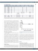
Table 2. Response outcomes.
Patient
Patient 1
Patient 2
Patient 3 Patient 4
Patient 5 Patient 6
Patient 7 Patient 8#
Patient 9*
Patient 10*
Patient 11* Patient 12*
Etiology Risk Treatment stratification days
EBV-HLH High 28
EBV-HLH low 28
EBV-HLH low 28 AIDs-HLH High 28
EBV-HLH High 28 EBV-HLH High 28
Unclear Low 28 CAEBV-HLH High 28
EBV-HLH High 5
Unclaer High 3
EBV-HLH High 7 AIDs-HLH Low 3
Reasons for discontinuation
/
/
/ /
/ /
/ /
Critically ill; Potential CNS involvement
Continuous progress
Progress after PR Continuous progress
Response from treatment to day28
CR
CR
CR CR
CR CR
CR Relapse after CR
HLH improvement
NR
PR
NR
Duration of Follow-up response until treatment
data cutoff (days)
Survival
353 / Yes
368 / Yes
264 / Yes 273 / Yes
237 / Yes 258 / Yes
222 / Yes
28 5
1
7
1
HLH-1994 No HLH-1994 Yes
HLH-1994 Yes
HLH-1994 Yes
HLH-1994 Yes
HLH: hemophagocytic lymphohistiocytosis; CR: complete response;PR: partial response;NR: no response; CNS: central nervous system; #: patients who were treated by HLH-1994 regimen after Ruxolitinib discontinuation, but reactivation occurred at the 4th week, and refused salvage treatment due to financial difficulty. *: patients who were treated by HLH- 1994 regimen after Ruxolitinib discontinuation, achieved complete remission, and stopped treatment after 8 weeks except patient-12 who still underwent maintenance treatment.
no fever (body temperature <37.5°C), no cytopenia (absolute neu- trophil count ≥1.0×10⁹/L and platelet count ≥100×109/L with the absence of granulocyte colony-stimulating factor (G-CSF) and transfusion support must be documented for ≥4 days), no evi- dence of coagulopathy (fibrinogen levels >1.50 g/L), normal levels of soluble CD25, ferritin and triglyceride, normal spleen size as measured by abdominal ultrasound, and no neurological and CSF abnormalities attributed to HLH. A partial response was defined as normalization of ≥3 of the aforementioned HLH abnormalities (including CNS abnormalities) and no progression of other aspects of HLH disease pathology. HLH improvement was defined as at least a 50% improvement in ≥3 HLH abnormalities from baseline. At least a 50% worsening in two or more signs or laboratory abnormalities was considered progressive disease. Three or more symptoms and laboratory markers developing into abnormalities after achieving complete response was defined as relapse.
Side effects and complications
The safety population included all patients who received at least one dose of RUX.
Adverse events were assessed according to the National Cancer Institute Common Terminology Criteria for Adverse Events, ver- sion 3.0. (http://ctep.cancer.gov/protocol Development/electron- ic_applications/docs/ctcaev3.pdf). In detail, we monitored the enrolled patients for any signs of toxicity and complications every day for the first week of treatment, weekly during weeks 2-4, and monthly thereafter, including routine blood tests, coagulation, infections (i.e., latent tuberculosis, adenovirus, Epstein-Barr virus, cytomegalovirus, herpes zoster, Pneumocystis jirovecii and fungal infections), cardiac function (myocardial enzyme spectrum, elec- trocardiogram, ultrasound cardiogram), renal function (serum cre- atinine, urea nitrogen, and creatinine clearance), liver function (ALT, AST, GGT, ALB, TBIL, I-BIL, D-BIL), and other adverse drug reactions such as dizziness, headache, rash, dyspnea and gastroin- testinal reaction.
Statistical analysis
SPSS 20.0 software (SPSS, Chicago, IL, USA) was used for sta- tistical analysis. All normally distributed data are represented as means ± standard deviations, and comparisons of multiple param- eters between groups were performed by independent sample t-
Figure 1. Event-free survival of patients defined as the time from the initial dose of ruxolitinib to the first occurrence of disease progression, relapse or death (event).
tests. All data that were not distributed normally are represented as medians and ranges, and comparisons of multiple parameters between groups were performed by Wilcoxon rank sum tests. Patient survival was estimated by the Kaplan-Meier method, and differences in survival between groups were estimated by the log- rank test. In all analyses, P<0.05 was considered to denote a sig- nificant difference, and P<0.01 was considered very significant. For patients who discontinued RUX within 7 days, the last known values were used as the day 7 laboratory results.
Results
Characteristics of the patients
Twelve pediatric patients with newly diagnosed HLH were enrolled at Beijing Children’s Hospital from June 2019 to October 2019, including eight males and four females. The main clinical features of the enrolled patients at diag- nosis are summarized in Table 1. The median age was 4.7 (range, 1.3-13.4) years. The median duration before HLH
haematologica | 2021; 106(7)
1895
