
Page 117 - 2021_07-Haematologica-web
P. 117
Thrombopoietin maintains megakaryopoietic HSPC
levels of statistical significance are represented by asterisks. *P<0.05, **P<0.01, ***P<0.001. NS represents P>0.05.
Results
Hematopoietic stem and progenitor cells in bone mar- row require thrombopoietin from liver
To investigate the role of THPO in HSPC we first sought the source of THPO in the bone marrow. We gen- erated a Thpoflox mouse using CRISPR/Cas9 to simultane- ously insert two loxP sites flanking exons 2 and 3 of the Thpo gene (Figure 1A). Loss of Thpo mRNA expression in the liver of ThpoΔ/Δ Alb-Cre mice was confirmed by quantita- tive polymerase chain reaction analysis (Figure 1B). In agreement with Decker et al.11 we found that liver-specific knockout (KO) of THPO caused similar loss of platelets to that of Thpo-/-, while conditional KO models for megakaryo-cytes, HSPCs and bone marrow stromal cells (PF4, Vav and Osx respectively) did not show any signifi- cant difference (Figure 1D-H).11 Similarly, the numbers of bone marrow MkP were significantly reduced in ThpoΔ/Δ Alb-Cre mice and Thpo-/- mice, but not in other conditional KO models (Figure 1I-M). We also observed a similar pat- tern in the CD34-Flt3-LSK HSC population, with a reduc- tion in cell numbers within the bone marrow of ThpoΔ/Δ Alb- Cre mice similar to that of Thpo-/- mice, but no reduction in bone marrow-specific conditional KO models (Figure 1N-
AB
R). This would suggest that steady-state HSPC in the bone marrow mainly depend on THPO from the liver.
Thromobopoietin maintains megakaryopoietic progeni- tor numbers
We set out to find which other hematopoietic cells are influenced by THPO. Analysis of mature blood lineages revealed that B cells in the bone marrow (Figure 2A and B), T cells in thymus (Figure 2C and D) and white blood cells in peripheral blood (Figure 2E and F) were unaffect- ed by loss of THPO in both Thpofl/flAlb-Cre and Thpo-/- mod- els. No effect on peripheral blood red blood cells was seen in Thpofl/flAlb-Cre mice and although statistical analysis showed a significant reduction in Thpo-/- mice, this appears to be very low and may simply be an artefact of variance within the mouse strain (Figure 2G and H). To confirm that the loss of THPO did not affect mature lin- eages other than megakaryocytes, progenitor popula- tions in bone marrow were analyzed. Common lym- phoid progenitors in the bone marrow were unaffected by loss of THPO in both models (Figure 2I and J). Analysis of myeloid progenitors showed that only the previously defined Pre-GMP and Pre-MegE populations were affected by the loss of THPO (Figure 2K and L). Interestingly, these populations were previously shown to give rise to megakaryocytes in vitro while the other myeloid progenitors did not show megakaryopoietic potential,12 suggesting that only the MkP, Pre-GMP and
CD
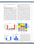
Figure 4. CD150- hematopoietic stem and progenitor cells have reduced thrombopoietin dependence and low megakaryopoietic potential. (A) Single cells of each hematopoietic stem and progenitor cell (HSPC) population were cultured in vitro with stem cell factor (SCF) or SCF and thrombopoietin (THPO) and colonies were counted on day 10. Bars show the percentage of sorted cells that gave rise to colonies (n=72 single cells across 3 experiments). (B) Single cells of each HSPC population were cultured in vitro and colonies analyzed by fluorescence activated cell sorting on day 10. Bars show the percentage of colonies that contain specific lineages (n=72 single cells across 3 experiments). (C, D) mRNA expression of Vwf (C) and Gfi1 (D) relative to GAPDH in HSPC (n=3). LT-HSC: long-term hematopoietic stem cell, Mk: megakaryocyte; MkE: megakaryocyte-erythroid; MkEGM: megakaryocyte-erythroid-granulocyte-macrophage; GM: granu- locyte-macrophage.
haematologica | 2021; 106(7)
1887
