
Page 114 - 2021_07-Haematologica-web
P. 114
A. O’Neill et al.
ground in accordance with Institutional Animal Care and Use Committee protocols. Alb-Cre mice in a white background were kindly provided by Prof. Qingde Wang of the University of Pittsburgh Medical Center and were back-crossed with C57BL/6-NTac mice at least five times.
Bone marrow mononuclear cell isolation
Mice were sacrificed by CO2 asphyxiation and femora, tibiae, pelves, humeri and vertebrae were crushed in phosphate- buffered saline (PBS). Red blood cells were lysed with NH4Cl in PBS and bone marrow mononuclear cells (BMMNC) were stained as described below.
A B
C
DEFGH
IJKLM
NOPQR
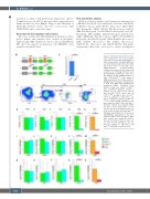
Figure 1. Thrombopoietin from liver main- tains hematopoietic stem and progenitor cells in the bone marrow. (A) Schematic of Thpo flox generation. Two guide RNA were used to simultaneously cut two sites flank- ing exons 2 and 3 of the Thpo gene. Single-stranded deoxyribo-oligonu- cleotides were then used to insert loxP sequences by homology directed repair. (B) Quantitative polymerase chain reac- tion analysis of Thpo mRNA from livers of Thpofl/fl and ThpoΔ/Δ Alb-Cre mice relative to B2M expression (n=3). (C) Flow cytometric gating system for analysis of hematopoiet- ic stem cells (HSC) and megakaryocyte progenitors (MkP). Peripheral blood platelet counts were taken for ThpoΔ/Δ mice and Thpofl/fl littermate controls of PF4-Cre mice (D, n=5fl/fl /6Δ/Δ), Vav-Cre mice (E, n=5fl/fl /6Δ/Δ) Osx-Cre mice (F, n=5fl/fl /6Δ/Δ) and Alb-Cre mice (G, n=5fl/fl /6Δ/Δ) in addition to Thpo-/-mice and ThpoWT/WT littermate controls (H, n=4WT/WT/6- /-). Cell counts of MkP population in the bone marrow of ThpoΔ/Δ mice and Thpofl/fl littermate controls were analyzed in PF4- Cre mice (I, n=6), Vav-Cre mice (J, n=6), Osx-Cre mice (K, n=6) and Alb-Cre mice (L, n=6) as well as Thpo-/-mice and ThpoWT/WT littermate controls (M, n=6). Cell counts of CD34-flt3-HSC populations in the bone marrow of ThpoΔ/Δ mice and Thpofl/fl litter- mate controls were analyzed in PF4-Cre mice (N, n=6), Vav-Cre mice (O, n=6), Osx- Cre mice (P, n=6) and Alb-Cre mice (Q, n=6) in addition to Thpo-/-mice and ThpoWT/WT littermate controls (R, n=6).
Flow cytometric analysis
All flow cytometric analyses and sorting were performed on a BD FACS Aria II cell sorter. Antibodies used were CD4 (RM4- 5, BD Biosciences), CD8a (53-6.7, eBioscience), B220 (RA3- 6B2, Biolegend), CD11b (M1/70, BD Biosciences), Ly6G/C (RB6-8C5, Biolegend), Ter119 (TER-119, Biolegend), Sca1 (D7, Biolegend), MPL (AMM2, Immuno-Biological Laboratories), CD41 (MWReg30, BD Biosciences), CD150 (TC15-12F12.2, Biolegend), cKit (2B8, Biolegend), CD34 (RAM34, eBioscience), IL7R (A7R34, eBioscience), Flt3 (A2F10, eBioscience), CD16/32 (93, eBioscience) and CD105 (MJ7/18, Biolegend). Streptavidin (eBioscience) was used to resolve biotinylated
1884
haematologica | 2021; 106(7)
