
Page 10 - 2021_07-Haematologica-web
P. 10
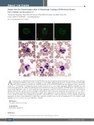
ABOUT THE COVER
Images from the Haematologica Atlas of Hematologic Cytology: MYH9-related disease Carlo L. Balduini1 and Alessandro Pecci2
1Ferrata-Storti Foundation, Pavia, Italy and 2University of Pavia-IRCCS Policlinico San Matteo, Pavia, Italy E-mail: CARLO L. BALDUINI - carlo.balduini@unipv.it
doi:10.3324/haematol.2021.279022
Acharacteristic of MYH9-related disease (MYH9-RD), the most frequent inherited thrombocytopenia, is the presence of aggregated MYH9 protein in the cytoplasm of neutrophils. Immunofluorescence staining of normal neutrophils shows the homogeneous distribution of MYH9 protein (A). In MYH9-RD MYH9 protein aggregates due to mutations in the N- or C-terminus. C-terminal mutations usually result in one or a few large aggregates which potentially associate with small aggregates (B), while N-terminal mutations (mutations in the motor domain) of MYH9 lead to the formation of many small aggregates only (C). Both small and large aggregates, named Döhle-like inclusion bodies, are detectable by immunofluorescence analysis in all neutrophils of all patients with a detection specificity and sensitivity close to 100%. However, in May-Grünwald-Giemsa stained blood films, these aggregates are more difficult to identify and are only detect- ed in a small percentage of neutrophils in around 50% of patients. Due to their characteristic round or spindle shape large aggregates are more easily identified as light blue corpuscles usually located at the cell periphery (D to F), whereas small aggregates (G to I, arrows) are more difficult to identify.1
Disclosures
No conflicts of interest to disclose.
Contributions
Both authors contributed.
Reference
1. Balduini CL, Pecci A. Inherited thrombocytopenias. Haematologica. 2020;105(Suppl 1):S237-247.
1780
haematologica | 2021; 106(7)
