
Page 9 - 2021_06-Haematologica-web
P. 9
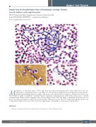
Images from the Haematologica Atlas of Hematologic Cytology: filariasis
Antonello Malfitano1 and Rosangela Invernizzi2
1IRCCS Policlinico San Matteo Foundation and 2University of Pavia, Pavia, Italy E-mail: ROSANGELA INVERNIZZI - rosangela.invernizzi@unipv.it
doi:10.3324/haematol.2021.278713
Microfilariae, i.e. the larval stage of Filaria spp., may be found in the peripheral blood after white blood cell con- centration by centrifugation and, occasionally, also in bone marrow preparations and other fine-needle aspirates. Morphological findings differ according to the species and form the basis for diagnosis. In buffy coat smears, Loa loa microfilariae appear as primitive serpentine-shaped organisms containing many nuclei with a head space, a sheath unstained with Giemsa and the tapering of the tail (top image). In the lower left image, a thick film shows the size of a Loa loa microfilaria in relation to white blood cells and the coiled tail; note also the row of nuclei through the whole body of the parasite right to the end of the tail (bottom right image).1 Eosinohilia is often an associated feature.
Reference
1. Malfitano A, Invernizzi R. Parasitic and fungal diseases. Haematologica. 2020; 105(Suppl. 1):29-39.
ABOUT THE COVER
haematologica | 2021; 106(6)
1511
