
Page 165 - 2021_06-Haematologica-web
P. 165
E4 enhances human HSPC engraftment in NSG mice
the presence of endogenous estrogens in these female ani- mals. Future experiments should be done with ovariec- tomized mice or taking the estrous cycle of female recipi- ents into account to identify the real effect of estrogen treatment on HSPC in female recipients. Furthermore, E4 treatment enlarged the hCD34+ cell population in already boosted human hematopoietic engraftment, but it had
less impact on the hCD34+hCD38- cell population and on secondary transplant (Figure 5D and E). Nevertheless, although the percentage of hCD34+hCD38- cells was unmodified by estrogen treatment, the total cell number of this primitive population was increased since human engraftment was higher in estrogen-treated mice (Figure 5B and D).
ABC
DEF
G
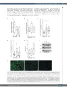
Figure 6. Estrogens modulate the hematopoietic niche. (A) Human engraftment in bone marrow (BM) of male mice transplanted with cells expanded from an initial dose of 5x10⁴ hCB-CD34+ cells after 4 days in culture in the presence of 100 nM E2 or E4. Human engraftment was analyzed 2 months after transplantation. (B) Human engraftment in BM of male mice transplanted with cells expanded from an initial dose of 5x10⁴ hCB-CD34+ cells after 1 week in co-culture with irradiated human BM-mesenchymal stromal cells (MSC) in the presence of 100 nM E2 or E4. Human engraftment was analyzed 3 months after transplantation. (C) Relative percentage of mouse MSC (mCD45-Ter119-hCD45-hCD235a-mCD140a+) in the BM of male mice transplanted with 5x10⁴ hCB-CD34+ cells, analyzed 4 months after transplantation. (D) Relative percentage of mouse vascular endothelial cells (mCD45-Ter119-hCD45-hCD235a-mCD144+) in the BM of the male mice transplanted with 5x10⁴ hCB-CD34+ cells, analyzed 4 months after transplantation. (E) Number of fibroblast colony-forming units (CFU-F) derived from the BM of vehicle- or estro- gen-treated mice after sublethal irradiation. (F) Representative agarose gel showing the quantitative real-time polymerase chain reaction products of ESR1 (top panel), ESR2 (middle panel) and HPRT1 (bottom panel) in human BM-MSC. (G) Representative immunofluorescence image of human BM-MSC stained with anti- ESR1 (green, left panel), anti-ESR2 (green, middle panel) or secondary antibody (green, right panel) and DAPI (blue). Data were obtained from three biological repli- cates and are presented by dots and box-plots that represent the interquartile range (p75, upper edge; p25, lower edge; p50, midline; p95, line above the box; and p5, line below the box). Statistical significance was analyzed by the Mann-Whitney U test: **P<0.01.
haematologica | 2021; 106(6)
1667
