
Page 9 - 2021_05-Haematologica-web
P. 9
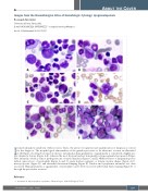
Images from the Haematologica Atlas of Hematologic Cytology: dysgranulopoiesis
Rosangela Invernizzi
University of Pavia, Pavia, Italy
E-mail: ROSANGELA INVERNIZZI - rosangela.invernizzi@unipv.it
doi:10.3324/haematol.2021.278517
In myelodysplastic syndrome without excess blasts, the precise recognition and quantification of dysplasia is critical for the diagnosis. The morphological abnormalities of the granulocytic series to be taken into account are illustrated in the Figure, showing bone marrow smears. Cytoplasmic hypogranularity or agranularity is considered a highly spe- cific dysplastic feature (Figure A-E), whereas the most frequent nuclear abnormality is hyposegmentation (pseudo-Pelger- Huët anomaly), which is almost pathognomonic of myelodysplasia (Figure C and E). Other features of dysgranulopoiesis include anisocytosis of neutrophils (Figure A and C), giant nuclear segments or bizarre nuclear shapes (Figure A-C), macropolycytes (Figure D), and abnormal chromatin clumping (Figure E). Nuclear and cytoplasmic anomalies are often associated. Hypogranularity or agranularity of neutrophils can be better assessed with Sudan black staining (Figure F) or through the peroxidase reaction.1
Reference
ABOUT THE COVER
1. Invernizzi R. Myelodysplastic syndromes. Haematologica. 2020;105(Suppl 1):78-97.
haematologica | 2021; 106(5)
1229
