
Page 289 - 2021_05-Haematologica-web
P. 289
Case Reports
being diagnosed with lethal hydrops fetalis. The birth weight of II1 was 1.9 kg, which is approximately the same as a normal child born at 34 weeks. The Apgar scores were normal and a physical examination showed that he had no congenital abnormalities, except for hydrocele. He was cared for as a premature infant in the neonatal department for 16 days. He was not transfused for the first 24 hours and did not enter the intensive care unit. The full postpartum clinical characteristics of the twins are summarized in the Online Supplementary Table S1.
The II1 received regular blood transfusions once a month from the age of 14 days due to anemia, and the anemia countenance and splenomegaly was comparable to a 1.5-year old. He grew normally, weighed 12 kg and was 85 cm in height at the age of 2 years. This matches the data of normal peers in southern China. He also had
normal intellect, in accordance with the evaluation stan- dard of the Chinese population.9
In order to clarify the reasons for the difference between the twins, we performed molecular analysis. Preliminary thalassemia mutation analysis in a local hos- pital showed that the parents were both heterozygous and II1 was homozygous for the SEA deletion without b-globin gene mutations. The II2 twin was not tested due to lack of sample, but his symptoms indicated that he was also likely to be homozygous for the SEA deletion. However, in our laboratory, a residual b-globin gene was detected in the blood cells of II1, based on the results of gap-ploymerase chain reaction (gap-PCR) and multiplex ligation-dependent probe amplification (MLPA) analyses (Figure 1D). This residual b-globin gene was also found in II1’s blood, hair follicle and oral mucosa cells with a dif- ferent level (2.58±0.64%, 8.91±0.65% and 3.58±0.49%,
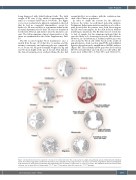
Figure 3. A hypothetical model to explain the etiology of the chimerism in II1. Two oocytes arise in a single zona pellucida and are fertilized by two individual sperm. The fertilized eggs divide and then the blastomeres fuse during the morula stage. Two inner cell masses are formed along with the division and migration of the blastomeres in the blastocyst (whether the trophoblastic layer is chimeric is unclear due to a lack of research). Two individuals then form from the inner cell mass, one of which (II1) is a chimera with --SEA/--SEA and --SEA/αα and the other (II2) carries the --SEA/--SEA genotype.
haematologica | 2021; 106(5)
1509
