
Page 238 - 2021_05-Haematologica-web
P. 238
Letters to the Editor
A
BC
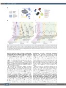
Figure 1. Immune response in COVID-19 patients and hematological cancer patients. (A) Schematic representation of semi-automated analysis of flow cyto- metric data and clustering of 17 immune cell types systematically identified in a total of 513 patients diagnosed with coronavirus disease 2019 (COVID-19). (B) Relative distribution of immune cell types in COVID-19 patients with (n=10, dots) and without (n=503, boxes) blood cancer. (C) Ratio between the percentage of each immune cell type at last follow-up and presentation in COVID-19 patients with (n=6, dots) and without (n=161, line) hematological tumor. Blue dots indi- cate alive patients and red dots represent deceased patients. Dashed line represents no variation over time (ratio = 1).*P<0.05; **, P<0.01; ***P<0.001. PC: plasma cells. : NK: natural killer cells.
immune cell types in COVID-19 patients bearing a hema- tological tumor when compared to other cases. Although the immune kinetics were quite variable, cancer patients dying from COVID-19 tended to have increased numbers of neutrophils counterbalanced by reduced percentages of other immune cell types versus those who survived (Figure 1C). Additional studies in larger series are war- ranted to confirm these findings; if so, individualized management of COVID-19 patients with hematological tumors according to their immune status at presentation and during follow-up should be investigated to improve outcomes in this population.
In an attempt to confirm these findings in a larger series, we were able to analyze PB samples from five additional COVID-19 cases with blood cancer from other hospitals who also performed immune profiling by MFC. A detailed analysis of major immune cell types in the 15 hematological cases versus the 503 COVID-19 patients without blood cancer, confirmed a recurrent altered dis- tribution of basophils, eosinophils, neutrophils, mono- cytes, natural killer (NK), T and B lymphocytes as well as circulating plasma cells (PC) (Figure 2). Furthermore, such alterations appeared to be more profound in deceased cases (two with acute myeloid leukemia and two with lymphoma) and reached statistical significance regarding B-cell distribution. Humoral immunity is critical for the clearance of SARS-CoV-2, as evidenced by the rapid and
near-universal detection of virus-specific neutralizing antibodies. Thus, very low B-cell numbers in patients with blood cancer due to tumor expansion and/or specif- ic drugs (e.g., immuno[chemo]therapies targeting B-cell and PC antigens) could emerge as another biomarker to predict disease severity after SARS-CoV-2 infection, together with advanced age and comorbidities that com- monly affect hematological cases. Noteworthy, the medi- an age of COVID-19 patients with or without blood can- cer was 73 and 60 years, respectively (P=0.049), which could also have contributed to poorer outcomes in the former.
SARS-CoV-2 infection of respiratory epithelial cells has been shown to activate monocytes, macrophages and dendritic cells,11 with an increasing number of studies suggesting that heightened inflammation is a defining feature of severe COVID-19. Thus, we specifically aimed at comparing the transcriptional profile of myeloid and antigen-presenting cells in COVID-19 patients with (n=3) or without (n=10) a hematological tumor. Unsupervised hierarchical analysis of RNAseq data from basophils, myeloid and plasmacytoid dendritic cells, classical and non-classical monocytes and neutrophils showed consid- erable clustering of samples from hematological cases (Online Supplemental Figure S3A). Furthermore, a variable number of differentially expressed genes was found in all six cell types between COVID-19 patients with or with-
1458
haematologica | 2021; 106(5)
