
Page 18 - 2021_05-Haematologica-web
P. 18
Fleur S. Peters et al.
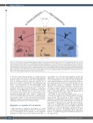
Figure 2. T-cell differentiation. The differentiation pathway of exhausted T cells (left panel) is distinct from that of effector T cells (right panel), and these cells show clear phenotypic and functional differences. The differentiation pathway of dysfunctional CLL T cells (middle panel) is currently unknown and these cells show fea- tures of exhausted as well as effector T cells. The dotted lines represent the possibility of CLL T cells to be on the path towards exhaustion or effector cell. The phe- notypic characteristics depicted here are cell-surface markers, metabolic features and transcription factor profiles. PD-1: programmed death protein-1, CTLA4: cyto- toxic T-lymphocyte-associated protein-4, TIM3: T-cell immunoglobulin and mucin-domain containing-3, TIGIT: T cell immunoreceptor with Ig and ITIM domains, KLRG1: killer cell lectin-like receptor subfamily G member-1, ROS: reactive oxygen species, IL-10: interleukin-10, TBG-b: transforming growth factor-beta, IFNγ: interferon- gamma, TNF: tumor necrosis factor, TCF-1: T cell factor-1, TOX: thymocyte selection-associated high mobility group box protein, T-bet: T-box expressed in T cells, Eomes: eomesodermin, Blimp-1: B-lymphocyte-induced maturation protein-1.
is clear that, rather than an analysis of a single transcrip- tion factor, the study of CLL T cells requires an integrated network analysis of genome-wide data. Integrating the level of transcription factor expression with the set of genes they control, could help uncover mechanisms that lead to T-cell dysfunction in CLL. Interestingly, recent studies demonstrated recovery of T-cell function upon treatment with Venetoclax and Ibrutinib,28,78 indicating a plasticity of T-cell phenotypes by therapeutic eradication of the tumor cells. Whether this recovery is due to selec- tive survival of functional T cells or true reversibility of the dysfunctional phenotype, due to return to normal home- ostasis, is currently unknown. We believe analyzing epige- netic profiles of these T cells could provide answers since epigenetic regulation can represent an interface between a changing environment and T-cell functionality.
Epigenetics as regulator of T-cell function
Tight regulation of epigenetic mechanisms is essential for immune cell differentiation (Table 1)22,79 and the func- tion of transcription factors depends on the epigenetic landscape in which they operate. Instructed by intra- or
extracellular cues, cells alter their epigenetic profile and differentiate, a process that is hierarchical; cells generally do not de-differentiate in vivo. Naive T cells are character- ized by high methylation and closed chromatin of effector genes and, upon activation, an accessible and gene activat- ing epigenetic profile is established.79 Promoters and enhancers often retain their permissive chromatin state, allowing for rapid re-activation of effector function upon re-encounter with antigen.79 This epigenetic memory helps to understand the cell-of-origin and lineage history of dysfunctional T cells found in cancer.
The CD4+ T cells in the CLL TME produce a set of cytokines that implicates their cellular identity and epige- nomic profile. The production of IFNγ, a typical Th1 cytokine, is strongly regulated by histone acetylation and DNA methylation. T-bet establishes these activating his- tone marks on the IFNG locus63 and Th1 cells are charac- terized by a demethylated IFNG promoter, allowing expression. Normally, Th1 cells retain highly methylated promoters of other lineage-specific cytokine genes, for example IL4 and IL17, preventing expression of these genes.80 This epigenetic programming allows differentia- tion into a specific lineage and concomitant exclusion of the opposite fate.
1238
haematologica | 2021; 106(5)
