
Page 107 - 2021_05-Haematologica-web
P. 107
Endothelial injury, F-actin and vitamin-D binding protein
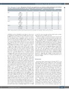
Table 4. Mononuclear cell analysis. Mononuclear cells from the same normal donor were incubated overnight in individual wells with individual sera from 18 children receiving hematopoietic stem cell transplant (HSCT), collected at days 0, 30, 60 and 100 after HSCT.
IL6 TNFα IL10
r2 P r2 P r2 P
Day 0
Day 30
Day 60
Day 100
VDBP 25-OH Vit D Free Vit D VDBP 25-OH Vit D Free Vit D VDBP 25-OH Vit D Free Vit D VDBP 25-OH Vit D
Free Vit D
0.14 0.58 0.063 0.80 0.077 0.76 -0.07 0.83 -0.32 0.28 -0.16 0.60
0.53 0.028 0.19 0.45 0.18 0.18
0.7 0.0013
0.47 0.05
-0.28 0.26
0.079 0.75 0.21 0.41 0.046 0.85 -0.27 0.37 -0.11 0.71 0.11 0.72 0.52 0.027 0.23 0.35 0.12 0.64 0.28 0.27 -0.12 0.63 -0.63 0.005
0.16 0.52
0.20 0.43 -0.061 0.81 0.10 0.73 0.36 0.23 0.21 0.48
-0.65 0.0036 -0.30 0.22 -0.16 0.53 -0.46 0.05 -0.40 0.10 0.24 0.33
Cytokine levels were measured by enzyme-linked immunosorbent assay in supernatant from each well.Cytokine levels were correlated with levels of vitamin D binding protein (VDBP), 25-hydroxy vitamin D (25-OH Vit D) and free vitamin D (Free Vit D); IL6; interleukin 6, IL10: interleukin 10; TNFα : tumor necrosis factor α.
(25-OH-D) and free 25-OH-D levels (Table 4). Data show no significant correlation between VDBP, and total or free 25-OH-D and any cytokine at days 0 and 30. In contrast, the day 60 data show a significant positive association between VDBP and IL-6 and TNFα, and a significant neg- ative association between VDBP and IL-10. The associa- tion between VDBP and IL-6 and IL-10 was also seen with sera collected at day 100. A positive association between 25-OH-D and IL-6 and a negative association between free 25-OH-D and TNFα was also observed at day 100.
These data suggest that serum VDBP and its cargo, 25-OH-D, have the potential to modify immune cell func- tion, but this effect was only evident at later time points following transplant. In an exploratory analysis, seeking direction as to the possible nature of this immune modu- lation we incubated PBMC from a normal volunteer overnight with serum from a person who had stable levels of VDBP at baseline and day 60 and with serum from a person in whom VDBP levels increased significantly at day 60 compared with baseline. Changes in the gene expression profile between the same cells conditioned with serum from these two distinct patients were com- pared using RNAseq, and the genes with the most notable change in expression are listed in the Online Supplementary Table S1. Observed changes in gene expression were con- sistent with altered macrophage function, consistent with the established potential for a deglycosylated form of VDBP to act as a macrophage activating factor. The RNAseq data, together with the change in the cytokine profile, supported a possible change in macrophage polar- ization as a consequence of exposure to VDBP after HSCT. We addressed this further by incubating CD14/16 expressing normal peripheral blood macrophages sepa- rately with serum collected from the same HSCT recipient at baseline (VDBP level 78) and at day 100 (VDBP level 907, genotype Gc1F/Gc1F) and from a second HSCT recip- ient in whom VDBP levels were more constant (baseline VDBP level 681, day 100 level 727, genotype Gc1S/Gc2). We then assessed macrophage polarization in the result- ing cell cultures by measuring the ECAR during a glucose stress test performed using Seahorse technology (Figure 3). Data indicate altered macrophage polarization in cells incubated with serum containing a higher level of VDBP
at day 100, but not in cells incubated with serum contain- ing lower levels of VDBP at day 100.
VDBP macrophage activating activity has been reported to vary by genotype and by post-translational glycosyla- tion.25-27 We hypothesized that changes in glycosylation of VDBP during the course of transplant might play a role in modifying macrophage polarization after HSCT. We test- ed this hypothesis by measuring glycosylation of VDBP in serum samples from two persons of differing VDBP geno- type, collected at days 0 and 100 after HSCT (four samples total) to see if there were significant changes in glycosyla- tion during recovery after HSCT (Online Supplementary Table S2). The only significant change in glycosylation between days 0 and 100 occurred at a single minor glycan (HexNAc(1)Hex(1)NeuAc(2)), with no significant change seen in the major glycosylated glycan (HexNAc(1)Hex(1) NeuAc(1)), suggesting that changes in glycosylation are likely not a major contributor to changes in immune mod- ulation by VDBP.
Discussion
Endothelial injury immediately following HSCT is an important initiator of later complications such as TA-TMA and GvHD, which together are major causes of morbidity and mortality after HSCT.1 Effector mechanisms leading to damage to the endothelium have not been described, although direct injury by chemotherapy and injury sec- ondary to viral reactivation are possible causes. In this study we show that release of the angiopathic molecule F- actin into the circulation is associated with TA-TMA, an immediate clinical consequence of endothelial damage. F- actin is typically not present in the circulation of healthy individuals who have low and relatively constant levels of cell turnover that do not exceed the capacity of the actin scavenger system for rapid removal of the harmful pro- tein.12,13 Rapid lysis of the entire hematopoietic system during conditioning therapy for HSCT overwhelms actin scavenging, at least in some cases, and allows F-actin to remain in the circulation and cause damage. We also found that F-actin was more likely to be detected after a myeloablative than a reduced intensity preparative regi-
haematologica | 2021; 106(5)
1327
