
Page 67 - 2021_04-Haematologica-web
P. 67
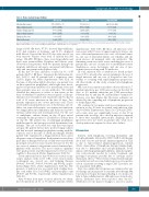
Cytopenias after CAR T-cell therapy
Table 3. Bone marrow biopsy findings Median Day (range)
Hypocellular marrow Normocellular marrow Hypercellular marrow Dysplasia Present
Fibrosis Present
Pre-axi-cel
-179 (-1551 to -7)
21·5% (14/65)
60·0% (39/65)
18·5% (12/65)
0·0% (0/65)
10·8% (7/65)
Day 1-100
31 (6 to 100)
87·5% (14/16)
12·5% (2/16)
0·0% (0/16)
6·3% (1/16)
31·3% (5/16)
Day 101-360
202 (102 to 289)
50·0% (3/6)
50·0% (3/6)
0·0% (0/6)
33·3% (2/6)
33·3% (2/6)
axi-cel: axicabtagene ciloleucel. Bone marrow cellularity as designated by the clinical pathologist. Clinical criteria account for patient age and are defined as hypocellular with a cell-to-fat ratio of 1:3 or less and hypercellular with a cell-to-fat ratio of 3:1 or greater.20
(range: 6-100). Of these, 87.5% showed hypocellularity, 6.3% had evidence of dysplasia, and 31.3% displayed mild fibrosis. Beyond the first 100 days after axi-cel, six bone marrow biopsies were done at median day 202 (range: 102-289). Of these, three were hypocellular and three were normocellular. Dysplasia and fibrosis were observed in four patients, with two patients having both dysplasia and fibrosis on biopsy, one patient with fibrosis only, and one patient with dysplasia only.
In the first 30 days, infection occurred in 31 of 85 patients (36.5%). Of these, 12 patients had Clostridium dif- ficile (14.1%), and 10 patients had a respiratory virus (11.8%) (Figure 3A, Online Supplementary Table S2A). At the time of Clostridium difficile detection, one patient was on fluoroquinolone prophylaxis, while eight were receiv- ing broad spectrum antibiotics for neutropenic fever and three patients were not on any antibiotics. We further classified the infections as severe or non-severe on the basis of whether IV antibiotics were required or if hospi- talization was warranted, and 11 patients (12.9%) had at total of 13 severe infections in the first 30 days. Two patients experienced two severe infections each. These severe infections included six cases of bacteremia, one cel- lulitis, one acute cholecystitis, one urinary tract infection, two fungal infections (candidemia and fusariosis) and two viremias (adenovirus and cytomegalovirus). For the case of candidemia, cultures drawn on day 38 post axi-cel eventually became positive for Candida krusei (5 days later on day 43). The patient was on antifungal prophylaxis with fluconazole, and autopsy revealed disseminated can- didemia. The case of Fusarium was only discovered upon autopsy; the patient did not have positive fungal cultures and had received antifungal prophylaxis starting with flu- conazole, and at the time of death, micafungin. Neither patient who experienced a fungal infection had a prior allogeneic stem cell transplant. Both patients received five prior lines of therapy before axi-cel and were treated with prolonged steroids for neurotoxicity after axi-cel. For the two cases of viremia, both patients received antiviral pro- phylaxis with acyclovir. The most common organisms were gram positive and the fungal isolates were not sus- ceptible to fluconazole (Online Supplementary Table S3).
Severe infections before day 30 were associated on uni- variate analyses with severe CRS, severe neurotoxicity, tocilizumab use, steroid use, and bridging therapy (P=0.007, P=0.007, P=0.03, P=0.004, and P=0.02, respec- tively) (Figure 3B-C, Online Supplementary Table S4). The presence of infection was associated with a higher base- line total white blood cell (WBC) and neutrophil count (P=0.03 and P=0.02, respectively) (Online Supplementary Table S5).
After 30 days from time of axi-cel infusion, we identi- fied 32 infections in 31 of 70 (44.3%) patients (Online
Supplementary Table S2B). Of these, 25 infections were non-severe with 19 cases of upper respiratory viruses, one case of bacterial pneumonia, two cases of bacterial sinusi- tis, one case of Clostridium difficile, one cellulitis, and one groin abscess, all managed with oral antibiotics. The remaining seven cases were severe, including five cases of pneumonia, one case of sepsis due to methicillin-resistant Staphylococcus aureus bacteremia, and one case of neu- tropenic fever resolving with IV antibiotics.
In total, infection was a contributor to death in three cases (3.5%), all early after axi-cel, including both cases of fungal infection and one case of Streptococcus mitis bac- teremia co-occurring with rapid lymphoma progression. All other deaths on this study occurred after lymphoma progression.
On a rate basis, infection incidence decreased from 11.7 incident infections per 1,000 person-days in the first 30 days to 2.3 incident infections per 1,000 person-days between day 31 and day 90, and incidence continued to decrease over time (Figure 4A). However, estimates are affected by the competing risk of lymphoma progression or death (Figure 4B).
We evaluated if cytopenias and/or poor immune recon- stitution at day 30 were associated with infection after day 30 (Online Supplementary Figure S3). CD4 and CD8 T- cell counts were not significantly lower at day 30 in patients who would go on to develop infection compared to those who remained infection-free. Similarly, day 30 IgG levels were not lower in patients who went on to develop infection compared to those who did not.
Discussion
Patients with lymphoma receiving fludarabine and cyclophosphamide followed by CD19 CAR T-cell therapy are at risk of acute and chronic immunosuppression. This is due to tumor-related immunosuppression, residual effects of prior lines of lymphoma therapy, lymphodeple- tion due to fludarabine and cyclophosphamide, B-cell aplasia with hypogammaglobulinemia, immunosuppres- sive corticosteroids needed for toxicity treatment, and prolonged cytopenias possibly due to cytokine and/or chemokine effects.8-11 Here we have characterized immune reconstitution and associated infections in a retrospective cohort of patients receiving a single CAR T product, axi- cel, in patients with LBCL.
First, we confirm that cytopenias are common after axi- cel therapy and may persist for months after therapy. On bone marrow biopsy the typical finding is of a normocel- lular to hypocellular marrow, although cases of myelodys- plastic syndrome have been reported after axi-cel.12 Attribution of bone marrow changes to axi-cel is unclear
haematologica | 2021; 106(4)
983
