
Page 307 - 2021_04-Haematologica-web
P. 307
Case Reports
A
B
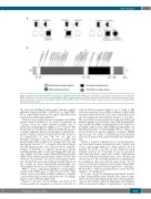
Figure 2. Pedigrees of the reported families and pathogenic variants described in the IARS2 gene. (A) Pedigrees, segregation and localization of the pathogenic IARS2 variants. (B) Linear map of the reported pathogenic IARS2 variants. Important structural domains and their locations on the protein map are shown. Amino acid location of each reported pathogenic variant is indicated (NM_018060.3). The likely pathogenic variants reported in this article are underlined and, if novel, indicated by a star.
the anticodon binding domain, and a missense variant inherited from his mother (c.2450G>A, p. Arg817His). The p. Arg817His variant has been previously reported in four patients with Leigh syndrome.5,8
Patient 2 was born from non-consanguineous healthy parents from Sri Lanka, at 31 weeks of gestation by cesarean section for cardiac rhythm anomalies after a dichorionic twin pregnancy (Apgar score 3, 6 and 7). His brother had no fetal distress and was healthy. He present- ed with respiratory distress and anemia at birth (hemo- globin 3.9 g/dL, normal 9-14 g/dL; MCV 84 fl). He need- ed transfusions at days 54, 69, 76 and 79. Thrombocytopenia was also present at birth (115 platelets/mm3 with a nadir at 50, normal >150.000) and myelogram showed 3.7% of ringed sideroblasts (Figure 1E). His plasma lactate was elevated (3.6-9.7 mmol/L, normal <2 mmol/L). A complete metabolic work-up, including plasma amino acids and vitamin B12 and uri- nary organic acids was normal. Brain MRI showed mild, diffuse white matter hyperintensities (Figure 1B). He was referred to the intensive care unit for mechanical ventila- tion. At day 54, he presented with pericardial effusion in the context of severe biventricular hypertrophic car- diomyopathy. He died at 2,5 months due to bradycardia.
Whole exome sequencing identified a homozygous variant in the IARS2 gene (c.199C>T; p. Pro67Ser). This variant, absent in publicly available databases, is highly conserved through evolution and predicted to be damag- ing by in silico softwares (Figure 1B).
Patient 3, the last of four siblings, was born at 33 weeks gestation by emergency caesarean section due to
reduced fetal movements (Apgar score 1, 5 and 6). His elder sister (patient 4, see pedigree in Figure 2) had a mild left intra-ventricular bleed on an early cranial ultrasound and was diagnosed with bilateral cataracts at 6 months. She was found to have global developmental delay and infantile spasms at 32 months of age with hypsarrhyth- mia on EEG. Her MRI neuroimaging showed volume loss involving caudate nuclei, globi pallidi and putamina, T2 flair hyperintensities of basal ganglia with no evidence of lactate doublet on nuclear magnetic resonance (NMR) spectroscopy. No cardiac or hematological involvement were noted and her bone marrow biopsy was unremark- able.
Patient 3 required ventilation for 2 weeks and devel- oped profound anemia (hemoglobin nadir 3.1g/dL) and severe cardiomegaly with poor cardiac function. At 4.5 months (11 weeks corrected), he was diagnosed with bilateral cataracts. At 18 months of age, he presented with a first metabolic crisis in the context of pneumonia associated with profound hypotonia, severe anemia and elevated lactate. He developed persistent oropharyngeal incoordination that necessitated gastrostomy. At 2.5 years, he developed refractory tonic seizures and an EEG demonstrated frequent bilateral spike-wave discharges. He was able to smile and acquired head control but was unable to sit unsupported.
At 4 years of age, the transfusion-dependent anaemia relapsed (hemoglobin nadir 3.5g/dL). Bone marrow aspi- ration showed subnormal myelopoiesis and megakary- opoiesis but marked erythroid hypoplasia and dysplasia with an increased number of progenitors, vacuolation
haematologica | 2021; 106(4)
1223
