
Page 301 - 2021_04-Haematologica-web
P. 301
Letters to the Editor
AB
CD
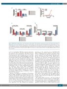
Figure 2. SRP68 biallelic variants induce a defect in granulocytic differentiation and endoplasmic reticulum stress and increase P53-dependent apoptosis of granulocytic precursors and neutrophils. CD34+ cells from the patient and the control donor were purified from blood and cultured for 12 days in serum free- medium with stem cell factor, interleukin 3 and granulocyte colony-stimulating factor. Sorted granulocytic cells CD33+15+11b– and CD33+15+11b+ were sorted on day 12. Expression levels were determined by quantitative everse transcription polymerase chain reaction related to PPIA and HPRT on day 12. (A) Expression level of SRP68 (n=1 in triplicate; mean±standard deviation [SD]). (B) Fold increase in cell proliferation during granulocytic differentiation (n=2; mean±SD). (C) Expression level of ATF4, CHOP and ratio spliced/unspliced XBP1 (n=1 in triplicate; mean±SD). (D) Expression level of P21, BAX, MDM2 and NOXA1 (n=1 in trip- licate; mean±SD).
tor (5 to 10 mg/kg/day). With this treatment and despite the persistent severe neutropenia, the patient has not presented severe bacterial infections. There was no famil- ial medical history. After excluding the genes classically involved in CN by targeted high throughput sequencing, we performed whole exome sequencing (WES) on a trio- based approach (Online Supplementary Appendix). WES diagnosed an intronic homozygous point substitution affecting the splice donor site of exon 1 (c.184+2T>C) of the SRP68 gene. This variant was detected at a heterozy- gous state in his mother and was absent in his father sug- gesting the presence in trans of a large deletion (Online Supplementary Table S1). Sanger sequencing and quantita- tive polymerase chain reaction (PCR) confirmed the splice site variant and the deletion of exon 1 of SRP68 (Figure 1B-C; Online Supplementary Table S2). This latter was inherited from his father confirming the compound heterozygous SRP68 genotype (c.184+2T>C) (exon 1 deletion) of the patient.
In order to determine the consequences of the intronic SRP68 variant on splicing, we performed reverse tran- scription PCR (RT-PCR) using primary fibroblasts from the patient, his parents, and a control donor. Agarose gel electrophoresis indicated that the patient and his mother harbor both the expected 213 bp fragment and a shorter product (176 bp). Sequencing of this fragment revealed the use of a predicted cryptic splice donor site located 37 bases upstream (c.147) and resulting in the premature
truncation of the mutated protein (Ala50Phefs*52) (Figure 1D). By western blot analysis, we found a drastic decrease of SRP68 protein expression in fibroblasts of the patient compared to his parents (Figure 1E) demonstrat- ing the loss-of-function effect of the SRP68 defects. Nevertheless, we observed a residual protein expression in the patient that may be due to a partial splicing defect as suggested by the semi-quantitative analysis of SRP68 transcripts in the mother (ratio 65%/35% for both 213 bp/176 bp fragments, Figure 1D). This finding was also consistent with the SRP68 transcript expression level determined from patient’s granulocytic cells. The residual expression of SRP68 could be explained by the specific post-transcriptional splicing mechanism related to GT>GC variants affecting the canonical 5’ donor splice site as identified in our patient (c.184+2T>C). In fact, 5′ splice site GT>GC variants may retain their ability to generate normal transcripts and be associated with a milder than expected clinical phenotype.8 SRP68 func- tions only as a heterodimeric structure with the SRP72 protein.9 We could speculate that null SRP68 variants would be lethal on the basis of what was recently shown in Srp72-/- mice.10
We purified CD34+ progenitor cells from peripheral blood and cultured them in serum-free medium supple- mented with stem cell factor (25 ng/mL), interleukin 3 (10 ng/mL) and granulocyte colony-stimulating factor (20 ng/mL) for 12 days. The level of SRP68 expression on day
haematologica | 2021; 106(4)
1217
