
Page 297 - 2021_04-Haematologica-web
P. 297
Letters to the Editor
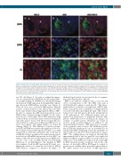
Figure 2. Immunofluorescent AID/BCL2 double staining. (A-C) Follicle (arrowhead) involved by in situ follicular neoplasia (ISFN) with BCL2 staining red cells (A) and few activation-induced cytidine deaminase (AID) staining green cells (B) shows lack of AID/BCL2 double staining cells( C). A nearby reactive follicle (arrow) is negative for BCL2 (A), positive for AID (B) and shows lack of AID/BCL2 double staining cells (C). (D-F) High magnification of the ISFN follicle showing BCL2 (D) positive cells (red), fewer AID (E) staining cells (green), and absence of AID/BCL2 double staining cells (F). (G-I) High magnification of a follicle from lymph node with FL: BCL2 (red) is diffusely positive in lymphoma cells (G) and AID (green) is positive in scattered lymphoma cells (H). AID/BCL2 double staining highlights many dual positive (arrows) lymphoma cells (I).
tive GC cells (Figure 1). In order to confirm this impres- sion, we then performed AID/BCL2 double immunofluo- rescent (IF) staining on 16 ISFN cases. Double IF staining showed lack of AID expression in strongly BCL2 express- ing cells in the ISFN follicles in all the cases (Figure 2). Thus, all cases were considered AID-negative. Internal positive controls (nearby reactive follicles and cells with- in the same follicle) stained appropriately (BCL2–, AID+). In order to compare the AID expression pattern seen in ISFN to that of typical systemic nodal FL, we also evalu- ated 15 cases of low grade FL for AID expression using double IF from a tissue microarray constructed during the same time period. Seven of 15 (47%) low grade FL cases demonstrated co-expression of BCL2 and AID in 10% or more of neoplastic cells within follicles on double IF staining (Figure 2). This difference between systemic nodal FL and ISFN was statistically significant (P<0.001). We could not evaluate AID expression in three cases with manifest FL at other time points/sites due to the lack of availability of these specimens. Given the known correla- tion between AID mRNA and protein levels by IHC,6 we further verified the lack of AID expression at mRNA level on two cases each of ISFN and FL by performing RNAscope in situ hybridization. The findings in ISFN were similar to both the IHC and double IF results, con- firming the absence (or extremely low levels) of AID in ISFN follicles, compared to manifest FL (Figure 3).
Methodological details are presented in the accompany- ing Online Supplementary Appendix.
ISFN is an indolent condition with a very low risk (<5%) of progression to FL.8 The ISFN cells carry a t(14;18)(q32;q21), similar to usual-type FL.9 This abnor- mality leads to constitutive overexpression of BCL2, inhi- bition of apoptosis and accumulation of inappropriately rescued B cells with a prolonged life span. This event is believed to be the first genetic hit in the natural history of FL pathogenesis, but additional genetic hits are required for progression to malignant follicular lym- phoma.10 ISFN carries few secondary genetic changes.9 In contrast, secondary genetic alterations are found in 70–90% of FL at initial diagnosis in addition to the t(14;18)/IGH-BCL2 fusion.11 AID expression is a marker of the germinal center reaction. The additional genetic hits in follicular lymphoma B cells are postulated to be facilitated by AID, contributing to genomic instability.10 A subset of FL cases (25-100%) has been shown to express AID.3,4,5 We confirm this in our study by showing that approximately half of the low grade FL cases expressed high level AID in neoplastic follicles by double IF. In our study, none of the ISFN cases expressed detectable AID in BCL2 intensely positive neoplastic cells in GC. The absence of detectable AID in IFSN may be related to greater genetic stability and its generally benign behavior. We cannot exclude very low levels of AID expression,
haematologica | 2021; 106(4)
1213
