
Page 257 - 2021_04-Haematologica-web
P. 257
Letters to the Editor
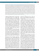
Figure 1. Effects of reveromycin A (RM-A) on osteoclasts (OC) and multiple myeloma (MM) cell viability. (A) Effects of RM-A on MM cell-bearing SCID-rab models. To prepare SCID-rab mice, rabbit femurs were cut into two and implanted subcutaneously in SCID mice. A month later the human MM cell line INA6 was inocu- lated directly into the bone marrow (BM) cavity in the rabbit bones implanted in SCID mice. After confirming the MM cell growth at 4 weeks after the MM cell inoculation, we started to inject RM-A at 4 mg/kg or a vehicle (saline) to the mice (n=5 for each treatment) intraperitoneally twice daily for 18 days. Soft X-ray and micro-computed tomography (mCT) images of the implanted rabbit femurs were taken before and after the treatment with RM-A or a vehicle. Representative images of soft X-ray (left panels) and mCT (right panels) are shown. MM tumor lesions are shown in red in 3D and cross sections of the rabbit bones in μCT images. (B) INA-6 cell-derived human soluble IL-6 receptor (sIL-6R) levels in mouse sera were measured as a marker for MM tumor burden after the treatment for 18 days with RM-A or a vehicle. The rabbit bones were taken out and analyzed to count the numbers of OC over bone surface (OC/bone surface). Data are expressed as the mean±standard error (SE). (C) Rabbit BM cells were cultured on the bovine bone slice in 96-well culture plates in RPMI1640 containing 10% fetal bovine serum with 20 ng/mL soluble receptor activator of NF-κB ligand (RANKL) for 4 days. After washing, RM-A at the indicated concentrations was added in triplicate for 24 hours (h) in the presence or absence of concanamycin A (CM-A) at 100 nM. The cells were then stained with tartrate-resistant acid phos- phatase (TRAP), and photos were taken (original magnitude, x200) (left). The numbers of TRAP-positive multinucleated cells (MNC) with three and more nuclei were counted (right). Data were expressed as % changes from the baseline (mean±SE). (D) INA-6, RPMI8226, OPM2 and MM.1S MM cell lines and primary MM cells were cultured at 2x10⁵/mL for 24 h at the indicated concentrations of RM-A. Viable cell numbers were counted with a WST-8 assay. The data were expressed as % changes from the baseline (mean±SE). (E) INA-6 cells were cultured in triplicate for 24 h in the presence of RM-A at the indicated concentrations with or without 5 mM metformin. Cell viability was analyzed by a WST-8 assay (left). The results were expressed as % changes from the baseline without any treatment (mean±SE. *P<0.05). Lactate levels in the culture supernatants were measured after the treatment with metformin for 24 h. (F) INA-6 and RPMI8226 MM cells were cultured in triplicate in the media whose pH values were adjusted by sodium hydroxide or lactic acid. RM-A was added at 1.0 μM. After culturing for 24 h, cell viability was analyzed by a WST-8 assay. Results were expressed as mean±SE. *P<0.05.
expanding in the BM with enhanced osteoclastogenesis. Under low O2 conditions, and as a consequence of gly- colysis (the Warburg effect), cancer cells highly produce protons and lactate, leading to an extracellular acidifica- tion to pH 6.4-7.0, while pH values are 7.2-7.4 in normal tissues.4 Activated OC on the bone surface abundantly secrete protons into excavated pits (~pH 4-5) to resorb bone while acidifying their close vicinity.5 In osteolytic bone lesions in MM, therefore, the MM cell-OC interac- tion appears to create a highly acidic milieu by protons produced by OC and lactate by proliferating glycolytic MM cells. We reported that acid activates the PI3K-Akt signaling to upregulate the acid sensor TRPV1 in MM cells, thereby forming a positive feedback loop between acid sensing and the PI3K-Akt survival signaling.6 In addi- tion, tumor acidity has been demonstrated to blunt cyto- toxic effects of various chemotherapeutic agents as well as the activity of immune effecter cells.7,8 Therefore, acidic conditions should be targeted to improve the ther-
apeutic efficacy against MM.
Reveromycin A (RM-A) is a small microbial metabolite
with three carboxylic groups, isolated from Streptomyces sp. SN-593.9,10 In an acidic microenvironment, RM-A becomes a non-polar form, which is able to permeate a cell membrane and induce apoptosis by inhibiting isoleucine tRNA synthesis.9,10 As such, RM-A has been demonstrated to preferentially induce apoptosis in acid- producing OC but not in other types of normal cells.9-11 In the present study, we explored whether RM-A targets an acidic condition induced by the MM cell-OC interaction to alleviate tumor expansion and bone destruction in MM.
To clarify anti-tumor activity of RM-A against MM, we examined the in vivo effects of RM-A in animal models mimicking MM bone lesions. The human MM cell line INA6 was inoculated into rabbit femurs subcutaneously implanted in SCID mice (SCID-rab), as previously report- ed.12 SCID-rab mice have been demonstrated to allow human MM cells to grow within the rabbit bones and induce bone destructive lesions as in patients with MM. In vehicle-treated mice, marked radiolucent osteolytic lesions were observed in the implanted rabbit bones on X-ray and micro-computed tomography (mCT) images, and MM tumor was packed in the BM cavity and expanded outside the rabbit bones (Figure 1A). However, in RM-A-treated mice, MM tumor markedly decreased in size without apparent bone destruction in rabbit bones. The levels of human soluble IL-6 receptor in mouse sera, a marker of human MM tumor burden, were also sub- stantially reduced in the RM-A-treated mice (Figure 1B). OC numbers were increased in bone specimens from
vehicle-treated SCID-rab mice; however, they were markedly reduced in RM-A-treated mice (Figure 1B). These results suggest that RM-A can suppress MM cell growth in the BM along with preventing bone destruc- tion and loss in vivo.
To further investigate the effects of RM-A, we first gen- erated OC on bone slices from whole rabbit BM cells, and then treated them with RM-A. Large multinucleated tartrate-resistant acid phosphatase (TRAP)-positive mature OC almost completely disappeared upon treat- ment with RM-A at 100 nM for 12 hours (h) (Figure 1C). Interestingly, blockade of acid release from OC by the proton pump inhibitor concanamycin A abolished the cytotoxic effect of RM-A on OC, indicating the critical role of acid released from OC in triggering the cytotoxic activity of RM-A. In contrast to OC, RM-A did not affect the viability of MM cell lines and primary MM cells even at higher concentrations up to 1 mM at 24 h (Figure 1D). However, RM-A was able to induce MM cell death even at concentrations as low as 100 nM when lactate produc- tion from MM cells was enhanced by metformin (Figure 1E). Furthermore, RM-A at 1 mM was able to induce cell death in MM cells when culture media were acidified to be at pH6.4 with exogenously added lactic acid (Figure 1F). These results suggest that acid-producing OC are highly susceptible to RM-A, and that an acidic milieu with lactate can trigger the cytocidal effects of RM-A against MM cells.
We next dissected the mechanisms of the MM cell death in acidic conditions by RM-A. RM-A induced apop- tosis in MM cells at pH6.4 but not at pH7.4, as indicated with annexin V-propidium iodine dual staining (Figure 2A). RM-A activated caspase-8 as well as caspase-9 in MM cells at pH6.4 (Figure 2B andC), indicating the induc- tion of caspase-dependent apoptosis. The transcription factor Sp1 has been demonstrated to be overexpressed and to act as a critical pro-survival mediator in MM cells.13,14 In parallel with the caspase-8 activation, the pro- tein levels of Sp1 were reduced in MM cells at pH6.4 but not at pH6.8 nor pH7.4 (Figure 2B). However, Sp1 mRNA was not decreased in MM cells even at pH6.4. We previ- ously reported that Sp1 protein is subject to enzymatic degradation by caspase-8, thereby inducing MM cell death.14 Consistent with the previous observation,14 treat- ment with the caspase-8 inhibitor z-IETD-FMK abolished the reduction of Sp1 protein at pH6.4 (Figure 2D), indi- cating caspase-8-mediated degradation of Sp1 protein. Furthermore, treatment with the Sp1 inhibitor terame- procol was able to reduce its target molecules critical for MM cell growth and survival, PIM215 and MYC14,15 (Figure 2E). Consistently, PIM2 and MYC levels were decreased
haematologica | 2021; 106(4)
1173
