
Page 242 - 2021_03-Haematologica-web
P. 242
Letters to the Editor
icantly lower than that in the discovery relapse cohort. The median time to relapse in all ZEB2mut-positive patients was 2.6 years from original diagnosis (range, 0.9- 5.3); the median follow-up time of those who remained in first continuous complete remission was 6.1 years (range, 1.7-10.2). Among 24 ZEB2mut-positive patients treated on frontline protocols utilizing minimal residual disease-based risk stratification, 7, 11 and 6 patients were stratified to standard, medium and high risk arms, respec-
tively.
Of the total 32 ZEB2mut patients identified within our
study (for patients’ characteristics including distribution into cohorts, see Table 1 and Online Supplementary Table S1), the ZEB2mut was already detected at the initial man- ifestation of leukemia in 29. Among these patients, we found a male to female ratio of 0.53, a median age at diagnosis of 8 years (10/29 patients were ≥10 years old) and a median initial white blood cell count of 8.2x10⁹/L (only 1 patient had a white blood cell count ≥50x109/L). The H1038R, Q1072R and Q1072K mutations were found in 14, 13 and 2 patients, respectively. At the DNA level, the diagnostic variant allele frequencies of ZEB2mut ranged from 2% to 79% (variant allele frequen- cy ≤20% in 12/25 cases), pointing to a frequent subclon- ality (and thus an unlikely primary origin of ZEB2mut). Variant allele frequencies of ZEB2mut alleles at the tran- script level were mostly higher or comparable to those at the DNA level. Among 14 analyzed patients with paired diagnostic/relapse samples, the ZEB2mut present at diag- nosis was preserved in ten and lost in one patient, while in three patients the ZEB2mut from relapse was unde- tectable at diagnosis and thus potentially newly gained.
We utilized RNA sequencing to investigate the pres- ence of subtype-defining genetic lesions, subtype-defin- ing gene-expression signatures4,6-14 and other known genetic lesions in ZEB2mut-positive ALL.
Of 32 ZEB2mut-positive ALL cases, six were classified as DUX4-rearranged; the TCF3-HLF fusion and ZNF384 rearrangement were found in one case each, while no subtype-defining lesion was detected by RNA sequenc- ing in 24 cases. Among these, iAMP21 was found by rou- tine cytogenetic investigation in one case, while it was not consistently investigated in all patients. ETV6- RUNX1-like and BCR-ABL1-like gene expression signa- tures were not detected in any patient (Online Supplementary Figure S1). Thus, the majority of ZEB2mut- positive cases (23/32; 72%) did not belong to any estab- lished ALL subtype.9
Variant and fusion analyses of RNA sequencing data revealed that additional genes/pathways were recurrent- ly affected in ZEB2mut-positive cases (Table 1). Of the 28 ZEB2mut cases analyzed by RNA sequencing at initial leukemia manifestation, seven had the P2RY8-CRLF2 fusion. Of note, this fusion was significantly enriched in cases with the Q1072 mutations compared to those with the H1038 mutation (47% vs. 0%, P=0.007). On the other hand, Ras pathway-activating mutations (muta- tions in NRAS, PTPN11 and FLT3) tended to be more fre- quent in cases with the H1038 mutation (38% vs. 7%, P=0.07). Interestingly, in addition to genetic differences, patients with the two ZEB2mut types also differed by age. Patients with an H1038 ZEB2mut were significantly older than those with a Q1072 ZEB2mut (median age 12.4 vs. 3.8 years; P=0.02). There was no significant dif- ference in white blood cell count and a modest trend towards a higher frequency of males in H1038 ZEB2mut- positive patients than in Q1072 ZEB2mut-positive ones (P=0.13).
888
haematologica | 2021; 106(3)
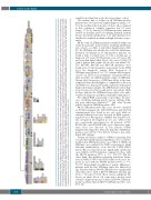
Figure 2. Unsupervised hierarchical clustering. Unsupervised hierarchical clustering based on the expression of 800 most differentially expressed genes (across the whole sample set, n =156) was performed using the ward.D method and Euclidean distance linkage. The figure shows the resultant dendrogram. The sample set includes samples from the patients with ZEB2-mutation-positive B-other acute lymphocytic leukemia (ALL) in the present study and samples from the patients with B-other ALL, BCR-ABL1-positive ALL and ETV6-RUNX1-positive ALL from our previous study.15 The BCR-ABL1-positive and ETV6-RUNX1-positive samples were included in the sample set to demonstrate the strength of co-clustering of ETV6-RUNX1-like and BCR-ABL1-like samples with their fusion-positive counterparts. The ETV6-RUNX1-like and BCR-ABL1-like samples were identified by supervised hierarchical clustering analyses (Online Supplementary Figure S1). The left-to-right order of samples can be found in Online Supplementary Table S4. ZEB2 H1038 and Q1072 mutation clusters are highlighted by red and blue boxes, respectively. DG: diagnosis; REL: relapse; r: rearrangement; low: variant allele frequency at the DNA level (or complementary DNA level if DNA not analyzed) <20%.
