
Page 225 - 2021_03-Haematologica-web
P. 225
Letters to the Editor
Clinical spectrum and therapeutic management of auto-immune myelofibrosis: a nation-wide study of 30 cases
In 2003, Pullarkat et al. described for the first time “pri- mary autoimmune myelofibrosis” (AIMF) as myelofibro- sis occurring in patients presenting autoimmune biologi- cal signs in the absence of a well-defined autoimmune disease (AID).1 Conversely, the term “secondary AIMF” is used for myelofibrosis occurring with a well-defined AID, most commonly systemic lupus erythematosus (SLE) but also systemic sclerosis, dermatomyositis, Sjögren syndrome,or organ-specific autoimmune diseases such as autoimmune hepatitis.
Being able to differentiate between an autoimmune or a clonal disease is crucial because of different therapeutic management, but can remains challenging. Since the dis- covery of gain of function (GOF) mutations of Janus kinase 2 (JAK2), novel findings such as calreticulin (CALR) GOF mutation and mutant myeloproliferative leukemia (MPL) protein have been described in clonal myelofibrosis. Interestingly, these findings seem to lack in case of autoimmune disease. Moreover, one of the fun- damental characteristics of AIMF seems to be its sensitiv- ity to glucocorticoids (GC),2 therefore GC remain the first-choice therapy. However, the long-term complica- tions of GC are severe, and other GC-sparing therapies should be considered. Unfortunately, little is known about the natural course of the disease and the optimal indications and efficacy of treatments.
The main goal of this study was to describe the presen- tation, the indications of current treatment and the course of 30 multicenter AIMF cases in France.
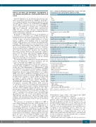
We performed a nation-wide, retrospective, and obser- vational study of AIMF by contacting two French net- works, the “Club Rhumatismes et Inflammation” (CRI) and “Maladies rares immuno-hématologiques” (MaRIH), dedicated to autoimmune diseases and rare immuno- hematologic diseases, respectively. Primary or secondary AIMF cases had to fulfill the following criteria to be included in the study: bone marrow (BM) fibrosis proven by BM biopsy, regardless of grade AND a defined AID according to current respective American College of Rheumatology/European League Against Rheumatism (ACR/EULAR) international classification criteria OR autoimmune cytopenia OR positive ANA detection (> 1:80 titers). Cases were excluded if the myelofibrosis could be explained by another condition (hematological disorder, solid neoplasia, chronic infection, toxic expo- sure known to induce myelofibrosis, radiotherapy, meta- bolic). All observations were reviewed by a steering com- mittee consisting of an internist and a rheumatologist specialized in the care of rare auto-immune disorders (TM and LA), and data were collected by using a stan- dardized and anonymized data form. Detailed clinical and biological characteristics of the 30 cases are shown in Table 1.
The presence of cytopenias at diagnosis of AID or onset during follow-up is not rare, and is mostly due to iron or vitamin deficiencies, chronic inflammation or autoimmune cytopenias. In our study, AIMF was diag- nosed during follow-up consultation for a known AID (mostly SLE and portosystemic shunts [pSS]) in 40% of cases, which strengthens the need to explore any new hematological abnormalities during follow-up and sug- gests the need for screening for AIMF at any time. Moreover, 50% of the cases for which geographic origin was available (22 of 30 patients) were of African or
Table 1. Clinical and biological characteristics of cases at the time of autoimmune bone marrow fibrosis diagnosis (n=30).
Characteristics
Sex Male
Female
Geographic origin (n=22)
European Afro-American North-African Asian
Age at diagnosis (years), median (IQR) AIMF
AID
Median delay between diagnosis of AIMF
and AID (years), median (IQR)
Known AID before AIMF onset Medianhemoglobinlevel(g/L)atAIMFdiagnosis,(IQR) 94(79–106)
6 (20%) 24 (80%)
9 (41%) 7 (32%) 4 (18%) 2 (9%)
37 (30–49) 31 (24–42)
0 (0–7)
12 (40%)
Median platelet count (109/L) at AIMF diagnosis, (IQR)
Median WBC count (109/L) at AIMF diagnosis, (IQR)
Leukocytes (n=30) Neutrophils (n=29) Lymphocytes (n=29)
Hypocomplementemia
Positive Coombs test (Coombs test availability n=25) Primary auto-immune myelofibrosis
Secondary auto-immune myelofibrosis
Associated reported AID
Systemic lupus erythematosus Primary Sjögren syndrome McDuffie vasculitis
Mixed connective tissue disorder Dermatomyositis
Immune thrombocytopenic purpura
Fibrosis grade in bone-marrow biopsy (n=28)
Grade 1 Grade 2 Grade 3
Available mutation status (n=11)
JAK2 negative
JAK2 and CALR negative
Triple negative (JAK2; MPL and CALR)
90.5 (75–182)
2.65 (1.8–4) 1.36 (0.85–2.17) 0.74 (0.5–1.1) 18 (60%)
13 (52%)
0 (0%)
30 (100%)
21 (70%) 5 (17%) 1 (3%) 1 (3%) 1 (3%) 1 (3%)
18 (64%) 9 (32%) 1 (4%)
7 1 3
AIMF: autoimmune myelofibrosis; AID: associated autoimmune disease; IQR: Interquartile range; WBC: white blood cell;
North-African origin, which could suggest that AIMF could be more frequent in these patients.
The presence of ANA is commonly described in patients with early-developing primary myelofibrosis4 but can be positive at titers of 1:80 in up to 13% of healthy individuals aged 21 to 60 year.5 Physicians should be aware of this important element as the presence of ANA does not necessarily indicate that myelofibrosis is secondary to an autoimmune process. Moreover, one should not neglect the possibility of having a true pri- mary myelofibrosis occurring during the course of an AID as some studies have described a 20% increased risk for the development of an myeloproliferative neoplasms in patients with auto-immunity.6
Mutational status was negative for all screened cases
haematologica | 2021; 106(3)
871
