
Page 195 - 2021_03-Haematologica-web
P. 195
DKK1 long peptide for myeloma immunotherapy
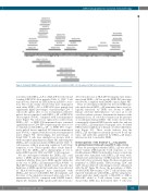
Figure 1. Dickkopf-1 (DKK1)3-76-long peptide (LP). Schematic presentation of DKK13-76-LP and epitopes for MHC class I and class II molecules.
four times with DKK13-76-LP or a HLA-DR*4-restricted and
detected in the sera of HLA-DR*4-transgenic mice immu-
nized with DKK1 -LP but not the DKK1-P30 short pep- 3-76
tide (P<0.01, compared with DMSO control) (Figure 4B).
Next, we determined whether the detected DKK1-spe-
cific antibodies in DKK13-76-LP-immunized mice were bio-
logically functional. As DKK1 was shown to inhibit
human osteoblast differentiation,20 we used an in vitro
osteoblast culture system to determine the function of the
immunized sera on osteoblast formation in the presence
of recombinant human DKK1. The results showed that
commercially obtained DKK1-specific antibodies and sera
from DKK1 -LP-immunized transgenic mice abolished 3-76
DKK1-induced inhibition of human osteoblast differentia- tion (Figure 4C). These results indicate that the DKK13-76-LP encompasses naturally processed B-cell epi- topes, and antibodies generated by DKK13-76-LP immu- nization are able to neutralize DKK1.
Immunogenicity of the Dickkopf-13-76-long peptide in priming human Dickkopf-1-specific T cells ex vivo
Next, we investigated whether DKK13-76-LP was able to induce human DKK1-specific T-cell responses ex vivo. Freshly prepared PBMC from healthy donors or myeloma patients were stimulated with DKK13-76-LP every week. The presence and frequency of DKK1-specific T cells were detected by flow cytometry. Figure 5A shows an increased frequency of DKK1-specific, IFN-g-secreting CD4+ and CD8+ cells in cultured T cells during in vitro (re)-stimulation with the LP. Figure 5B shows the percentages of HLA- A*0201-DKK1-P20 tetramer+ CD8+ T cells in cultures after repeated stimulations with DKK13-76-LP. By using mono- clonal antibodies (mAb) specific to HLA-DR or -DQ or HLA-ABC added to T-cell cultures before assay, we showed that MHC class II-restricted CD4+ T cells were
-binding DKK1-P30 short peptide (Table 1). CD4+ T-cell
response was detected by CFSE dilution and IFN-g secre-
tion. The results clearly showed that mice immunized
with either DKK13-76-LP or DKK1 P30 short peptide had
significantly higher percentages of proliferating CD4+ T
cells in the spleen after ex vivo re-stimulation with DC
pulsed, but not unpulsed, with DKK13-76-LP or DKK1-P30
short peptide (P<0.01, compared with non-immunized
mice) (Figure 3A). Moreover, splenocytes isolated from
+
DKK1 -LP - or DKK1 P30-immunized mice contained
3-76
significantly more CD4 IFN-g-expressing cells after ex vivo
re-stimulation with DKK13-76-LP - or DKK1 P30-, respec- tively, pulsed, but not unpulsed, DC than non-immunized mice (P<0.01, compared with those from non-immunized mice) (Figure 3B). Interestingly, the percentages of Foxp3+CD4+ T cells were similarly low in mice with or without peptide immunization (Figure 3B), indicating that DKK13-76-LP vaccination induced DKK1-specific CD4+ T- cell responses without promoting regulatory T-cell (Treg) formation in vivo. Taken together, these results demon- strate that the DKK13-76-LP is immunogenic in vivo to induce DKK1-specific CD4+ and CD8+ T-cell responses.
We also investigated whether the DKK13-76-LP could induce a DKK1-specific humoral immune response and examined whether there were DKK1-specific antibodies in the sera of DKK1 -LP-immunized HLA-A*0201- or
3-76
HLA-DR*4-transgenic mice. ELISA results showed that
high titers of DKK1-specific IgG antibodies were detected in HLA-A*0201-transgenic mice immunized with DKK13-76-LP, but not with DKK1 P20 short peptide, and the titers of the antibodies increased after each cycle of immunization (P<0.01, compared with DMSO control) (Figure 4A). Similarly, DKK1-specific antibodies were also
haematologica | 2021; 106(3)
841
