
Page 9 - 2021_02-Haematologica-web
P. 9
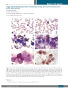
Images from the Haematologica Atlas of Hematologic Cytology: bone marrow metastases from gastric adenocarcinoma
Rosangela Invernizzi
University of Pavia, Pavia, Italy
E-mail: ROSANGELA INVERNIZZI - rosangela.invernizzi@unipv.it
doi:10.3324/haematol.2020.275529
In some patients with disseminated gastric adenocarcinoma, microangiopathic hemolytic anemia is the presenting fea- ture of the tumor. In this case of gastric adenocarcinoma with bone marrow metastases, an erythroblast, polychromat- ic red cells, microspherocytes and red cell fragmentation are observed in the peripheral blood smear (panels A and B). Bone marrow smear reveals a group of large tumor cells with irregular nuclear shape, and polychromatic, inhomogeneous, foamy or vacuolated cytoplasm (panels C and D). Periodic acid-Schiff (PAS) stain shows intense granular or diffuse cyto- plasmic positivity suggesting the accumulation of polysaccharides in the neoplastic cells (panel E). The strong immuno- cytochemical expression of cytokeratins (immunoperoxidase technique), in association with the absence of the common leukocyte antigen, confirms the non-hematopoietic origin of tumor cells (panel F).1
References
ABOUT THE COVER
1. Invernizzi R. Metastases of solid tumors. Haematologica. 2020; 105(Suppl 1):261-269.
haematologica | 2021; 106(2)
323
