
Page 74 - 2021_02-Haematologica-web
P. 74
P. Robach et al.
with rhEpo was progressive (Figure 2C). In contrast, trans- ferrin saturation (Tfsat) was not significantly altered by rhEpo injections (Figure 2D).
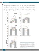
We also found concomitant increases in Epo and ERFE in healthy subjects exposed to a high altitude condition associated with O2 saturation of 85 ± 3% (Figure 4A, B). The correlation between ERFE and serum Epo found at high altitude (Fig 3B) and with rhEpo treatment (Figure 3A) suggests that hypoxia-related signaling is not directly
AB
involved in ERFE induction, as previously shown in mice.6 ERFE tended to increase more with micro-dose rhEpo than with exposure to high altitude (P=0.22), whereas Epo increased less with the micro-dose injections than with exposure to high altitude (P=0.04) (data not shown). We speculate that the shorter time of exposure to elevated Epo levels at high altitude (15 h) versus micro-dose injec- tions (24 h) may account for the observed trend. Tfsat, which was close to the level defining iron deficiency
CD
E
Figure 4. Effects of acute exposure to high altitude. Individual serum concen- trations and means ± standard deviations of erythropoietin (n=21) (A), ery- throferrone (ln transformed, n=21) (B), transferrin saturation (n=22) (C), hep- cidin (ln transformed, n=7) (D) and ferritin (n=22) (E), at sea level and after 15 h of exposure to high altitude (3800 m). Of note, out of 22 subjects, 15 sub- jects had sea-level hepcidin concentrations which were below the detection limit; therefore, the hepcidin statistical analysis was performed on seven sub- jects. P values denote differences between sea level and high altitude. Epo: erythropoietin; ERFE: erythroferrone.
388
haematologica | 2021; 106(2)
