
Page 335 - 2021_02-Haematologica-web
P. 335
Case Reports
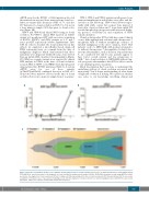
ddPCR assay for the RUNX1 c.292del mutation detected the mutation in unsorted bone marrow from clonal evo- lution at variant allele frequency (VAF) of 7% and also 497 days prior to clonal evolution at VAF <1%, but not in a bone marrow sample 2163 days prior to clonal evolu- tion (Figure 2B).
UPN-3 and UPN-4 had clinical NGS testing at clonal evolution. For UPN-3, clinical NGS reported an ASXL1 variant at 6 months post-IST with successive acquisition of RUNX1, SETBP1, and PHF6 variants at 2, 5, and 7 years post-IST respectively (Online Supplementary Table S1B). In order to elucidate the clonal architecture in this subject, we employed a microfluidics-based, single-cell DNA sequencing assay on a sample from the time of malignancy diagnosis which demonstrated that these mutations were all present within the same clone derived from an initial ASXL1 mutated clonal population (Figure 2C). While no somatic variants were reported by clinical NGS analysis in UPN-4 at the time of clonal evolution, research NGS of CD34+ sorted PBMC from this timepoint demonstrated two RUNX1 mutations (Table 1). A custom ddPCR assay for one of these variants (p.Thr257LeufsTer54) confirmed detection at VAF <2% in unsorted bone marrow collected at the time of clonal evolution but not in a marrow sample from 1 year prior.
UPN-1, UPN-2 and UPN-4 underwent allogeneic bone marrow transplantation and all three were alive and dis- ease-free at last follow-up. UPN-3 was followed expec- tantly with stable counts but a repeat bone marrow 2 years after clonal evolution and 7 years from initial IST revealed morphologic progression with increased dyspla- sia, presence of trisomy 21, and acquisition of PHF6 somatic mutation.
Clonal evolution after IST for SAA has a varied clinical course. Although high risk evolution with chromosome 7 aneuploidy with or without morphologic evidence of myeloid malignancy is the most common, fewer SAA patients evolve to MDS/AML with normal cytogenetics or without chromosome 7 abnormalities. Isolated chro- mosome abnormalities, such as deletion 13q and trisomy 8, are considered low risk because patients appear to have better overall survival and less progression to AML.11 Late clonal evolution to MDS/AML without typ- ical cytogenetic abnormalities after IST for SAA occurs in a very small proportion of patients.
Much investigation has been done to understand the underlying etiology of clonal evolution associated with monosomy 7 but a deeper understanding of other forms of high-risk evolution is lacking. We report here the first case series to our knowledge describing clinical and
C
AB
Figure 2. Detection of mutations in three severe aplastic anemia subjects prior to clonal evolution. (A) Presence of NPM1 mutation detected by digital droplet PCR (ddPCR) at, and 2 years prior to, the diagnosis of clonal evolution in unique patient number (UPN)-1; (B) RUNX1 variant was found using ddPCR at, and 1 year before, the diagnosis of clonal evolution in UPN-2; (C) single-cell DNA sequencing at the time of clonal evolution in UPN-3 demonstrates that the successive acquisition of mutations over a 7-year period, including 2 years after the diagnosis of clonal evolution to a myeloid malignancy were all within the same clonal population. SAA: severe aplastic anemia; VAF: variant allele frequency.
haematologica | 2021; 106(2)
649
