
Page 211 - 2021_02-Haematologica-web
P. 211
PolyP enhances uPA-mediated plasminogen activation
tions tested, however, the cofactor function of polyP was tempered at high concentrations. Inclusion of polyP delayed tPA-mediated lysis at all Glu-plasminogen con- centrations tested by an average of 40%; therefore, increasing the Glu-plasminogen concentration did not alter the efficacy of polyP on tPA-driven lysis.
Clots containing Lys-plasminogen lyse significantly faster than those containing Glu-plasminogen (Figure 5C-
D). A dose-dependent relationship exists between Lys-plasminogen concentration and uPA-mediated lysis, with 50 % lysis times decreasing from 51.1±1.36 min at 0.125 mM to 17.2±0.32 min at 1 mM (Figure 5C). In con- trast, there is no relationship between Lys-plasminogen concentration and tPA-mediated lysis, exemplifying the different modes of action of these plasminogen activators (Figure 5D). Similar to the results with plasmin generation,
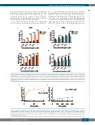
AB
CD
Figure 2. High concentrations of plasminogen attenuate the cofactor function of polyP in uPA-mediated plasminogen activation. Fibrin clots were prepared with
. Clotting was initiated with (5 mM). Plasmin generation in clots was quantified by incorporating the fluorogenic substrate D–VLK-AMC and monitoring fluores- cence release (FU; Ex 360 nm Em 460 nm). *P<0.05; **P<0.01, ***P<0.001 and ****P<0.0001 compared with control clots. Data are expressed as mean ± standard error of the mean, n≥3. polyP: polyphosphate; tPA: tissue plasminogen activator; uPA: urokinase plasminogen activator; FU: fluorescence units; Ex: excita-
2.4 mM fibrinogen, 0 -1 mM (A-B) Glu-plasminogen or (C-D) Lys-plasminogen, (A, C) 180 pM uPA or (B, D) 20 pM tPA ± 328 mM polyP
2
thrombin (0.25 U/mL) and CaCl tion; EM: emission.
65
AB
Figure 3. polyP binds to uPA with a significantly higher affinity than tPA. Binding of uPA or tPA (0–400 nM) to biotin-labelled polyP (71 mM) bound to streptavidin coated stripwells. Bound uPA (orange) or tPA (blue) was detected with chromogenic substrates (S2288 or CS-61 44 respectively) by reading the change in absorbance at 405 nm every 30 seconds (s) for 200 minutes (min). No unspecific binding was detected in the absence of biotin-labelled polyP (black lines). Data are expressed as baseline corrected nonlinear fit as mean ± standard error of the mean (SEM), n=4. PolyP: polyphosphate; tPA: tissue plasminogen activator; uPA: urokinase plasminogen activator.
haematologica | 2021; 106(2)
525
