
Page 204 - 2021_02-Haematologica-web
P. 204
S. Bobillo et al.
A
B
C D
E
F
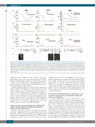
Figure 3. Cerebrospinal fluid (CSF) circulating tumor DNA (ctDNA) is more sensitive than flow cytometry (FC) to detect central nervous system (CNS) relapse and residual disease in CNS-restricted lymphomas. Longitudinal monitoring of three patients with CNS-restricted lymphomas (NHL3, NHL5 and NHL6) through (A) CSF and (D) plasma ctDNA analysis (variant allele frequency [VAF] values for each mutation obtained by droplet digital polymerase chain reaction [ddPCR]) and its corre- lation with (B) CSF flow cytometry (cells/mL), (C) cytology (+/- presence/absence of cells). (E) Type of treatment and response assessment, (F) radiological imaging (MRI). Data points are vertically aligned. MRI scans for patient NHL6 and NHL5 are not shown given that the patient only presented leptomeningeal involvement at diagnosis. Dx: time of diagnosis; IT MTX: intrathecal methotrexate; BAM-R: combined therapy (rituximab, carmustine, cytarabine and methotrexate); ASCT: autologous stem cell transplant; CR: complete response; LM: leptomeningeal; EOT: end of treatment; d: day; m: month; NA: not available. *CSF sample was not available for ctDNA analyses.
ctDNA is better identified in the CSF than in plasma. Interestingly, these two cases with restricted CNS lym- phoma in which plasma ctDNA was detected had a previ- ous history of systemic disease. This finding might reveal a systemic nature of some restricted SCNSL and deserves fur- ther investigation in larger studies.
We then analyzed 12 patients with systemic lymphoma without CNS involvement but with high risk for CNS relapse. In these cases, lumbar puncture was performed to rule out CNS infiltration and to administer IT MTX. As opposed to cases with CNS involvement, no ctDNA was detected in the CSF while it was detected at high VAF in the plasma of the majority of cases (Table 1). Finally, the patient with systemic and concomitant CNS disease had ctDNA in plasma but not in the CSF, suggesting that some CNS lesions cannot be captured through CSF ctDNA.
Central nervous system circulating tumor DNA exhibits higher sensitivity than flow cytometry in detecting central nervous system lesions
We next correlated the detection of ctDNA with the iden- tification of malignant cells by conventional tests in the same CSF samples at time of enrollment. Cytology detected the presence of malignant cells in 4 out of 6 CNS restricted lymphomas. In these four cases the FC analysis also detect-
ed lymphoma cells. In one case (NHL2), cytology was nor- mal but a small amount of tumor cells was observed by FC (0.06 cell/mL). Notably, all six cases exhibited CSF ctDNA. Importantly, in a patient with newly diagnosed PCNSL (NHL1) we could not detect lymphoma cells by FC or cytol- ogy; however, ctDNA was abundantly found in the CSF (Table 1). These results indicate that ctDNA analysis of the CSF can improve the detection of CNS lymphoma by con- ventional techniques.
Cerebrospinal fluid circulating tumor DNA can be used to monitor central nervous system tumor burden and response to treatment
To address whether the amount of CSF ctDNA could be useful to monitor tumor burden in CNS lymphomas, we analyzed sequential concomitant samples of CSF and plas- ma. CSF was obtained as part of the standard procedures before each administration of IT MTX or on suspicion of relapse. Tumor burden was evaluated by MRI and/or FC depending on the lymphoma location. We observed that CSF ctDNA levels correlated with disease response and progression (Figure 2). In addition, tumor burden measured by MRI and FC was concordant with CSF ctDNA levels in two patients with CNS lymphoma (Figure 2). Importantly, plasma ctDNA levels remained constantly low (VAF <6%)
518
haematologica | 2021; 106(2)
