
Page 201 - 2021_02-Haematologica-web
P. 201
ctDNA in cerebrospinal fluid of patients with CNS lymphoma
AB
C
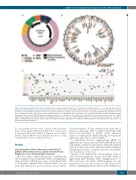
Figure 1. Genomic landscape of the cohort of study. (A) Pie chart representing the proportion of samples of each lymphoma type (see color legend). Two sectors in each sample depict whether the patient had systemic (gray) or central nervous system (CNS) restricted disease (black). (B) Circus plot representing all mutations identified across 16 lymphoma patients. A mutation was defined as driver according to Cancer Genome Interpreter (see Online Supplementary Appendix) colored in red (see Methods section). Genes with recurrent mutations have been highlighted in bold. (C) Bubble plot representing all driver mutations and droplet digital polymerase chain reaction (ddPCR) validated mutations identified in each patient. The bubble size represents the cancer allelic fraction (CAF) (see Online Supplementary Appendix) and the border has been highlighted in black if it has been found by ddPCR, in either the cerebrospinal fluid or plasma of each patient. NHL: non-Hodgkin lymphoma; DLBCL: diffuse large B-cell lymphoma; HGBCL: high-grade B-cell lymphoma; WM: Waldeström macroglobulinemia; MCL: mantle cell lymphoma; BL: Burkitt lymphoma.
the corresponding wild-type alleles. Genomic DNA from tumor tissues (10 ng), germline DNA from peripheral blood granulocytes (10 ng), plasma DNA, and CSF DNA (1-5 ng) were used for ddPCR analysis using the QX200 Droplet Digital PCR system according to manufacturer's protocols and the literature.20
Results
Circulating tumor DNA in the cerebrospinal fluid of patients with central nervous system restricted B-cell lymphoma is more abundant than in plasma
Here, we sought to analyze the presence of ctDNA in the CSF and plasma from 19 patients with B-cell lymphomas with and without CNS disease at time of enrollment (Table 1). Six patients exhibited CNS restricted disease, one sys- temic and CNS disease, and 12 systemic disease with no
CNS involvement. CSF was obtained at the same time as plasma in all patients. WES or targeted sequencing of the tumors was performed to identify somatic mutations (Figure 1). Variant-specific ddPCR detecting 1-5 variants per patient was performed to detect ctDNA.
To demonstrate that the variant-specific ddPCR was highly specific, we performed MYD88 L265P ddPCR in ten CSF samples obtained from patients with hydrocephalus without a brain tumor (n=6), and from patients diagnosed with glioma (n=3) and a cavernoma (n=1). No mutant allele was found in any of the cases.
Analysis of the ctDNA in the CSF and plasma of the six CNS restricted lymphomas showed detectable ctDNA at high variant allele frequency (VAF) (ranging from 1% to 95%) in all cases. In contrast, ctDNA in plasma was only detected in 2 out of 6 cases at very low VAF (always <5%) (Table 1), highlighting that in CNS restricted lymphomas,
haematologica | 2021; 106(2)
515
