
Page 100 - 2021_02-Haematologica-web
P. 100
M. Hu et al.
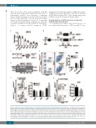
white blood cells, red blood cells and platelets in the PB, after miR-21 knockout (Online Supplementary Figure S1E, F). Interestingly, miR-21D/D mice exhibited a significant increase in the percentage of myeloid cells but a marked decrease in the percentage of B cells in the BM and PB (Figure 1E; Online Supplementary Figure S1G). Consistently, we found that the percentage of granulocyte-monocyte progenitors (GMP) was obviously enhanced, while the fre- quencies of common myeloid progenitors (CMP),
AB
megakaryocyte-erythroid progenitors (MEP) and common lymphoid progenitors (CLP) were reduced in the BM after miR-21 deletion (Figure 1F, G). These findings suggest that miR-21 is crucial for steady-state hematopoiesis.
Targeted deletion of miR-21 generates an aberrant hematopoietic stem cell pool
To determine how miR-21 affects hematopoiesis, we next analyzed the phenotypes of HSC from miR-21D/D mice
CE
D
FG
Figure 1. miR-21 is enriched in hematopoietic stem cells and its conditional ablation skews hematopoietic differentiation. (A) Quantitative real-time polymerase chain reaction (PCR) analysis of miR-21 expression in long-term hematopoietic stem cells (LT-HSC), short-term hematopoietic stem cells (ST-HSC), multipotent pro- genitors (MPP), common myeloid progenitors (CMP), common lymphoid progenitors (CLP), granulocyte-monocyte progenitors (GMP) and megakaryocyte-erythroid pro- genitors (MEP) isolated from normal 8-week old wild-type mice (n=4 mice). miR-21 expression was compared with that in LT-HSC. Gating strategies are provided in Online Supplementary Figure S1A, B. (B) The strategy for the generation of the conditional miR-21 knockout mouse model. For detailed genotyping see Online Supplementary Figure S1C. (C) Schematic for pIpC-inducible deletion of miR-21 in the hematopoietic system. (D) PCR-based analysis of genomic DNA from the bone marrow (BM) and spleen (Sp) of miR-21fl/fl and miR-21∆/∆ mice. (E) Flow cytometric analysis of the percentages of T cells (CD3e+), B cells (B220+) and myeloid cells (Gr-1+ and Mac-1+) in the BM of miR-21fl/fl and miR-21∆/∆ mice (n=6 mice per group). (F, G) Flow cytometric analysis of the percentages of (F) CMP, MEP, GMP and (G) CLP in the BM of miR-21fl/fl and miR-21∆/∆ mice (n=6 mice per group). All data are shown as means ± standard deviation. *P<0.05, **P<0.01.
414
haematologica | 2021; 106(2)
