
Page 87 - 2020_11-Haematologica-web
P. 87
TRPV4 mediates marrow adipocyte remodeling in AML
function of TRPV4 in BM adipocytes, respectively. 4aPDD is the first synthetic TRPV4 agonist and is a non- protein kinase C activated phorbol ester.20 Online Supplementary Figure S1A shows the half maximal inhibitory concentration (IC50) of the effects of RN1734 on BM adipocytes. Considering excessive Ca2+ influx could cause some toxicity to adipocytes,21 we aimed to find a concentration that has minimum cellular toxicity and promotes the Ca2+ influx needed for our experiments. As Online Supplementary Figure S1B shows, 4aPDD at a concentration of 0.25 mg/mL resulted in an acceptable level of toxicity of adipocytes, while allowing Ca2+ influx to reach the level required for the experiment. Oil red O staining and quantitative analysis showed that RN1734 reduced the number and area of BM adipocytes, whereas 4aPDD did not induce a similar change (Figure 1C, D), suggesting that the inhibition of TRPV4 contributes to reducing BM adipocyte number and size. Furthermore, optical density value measurements showed that lipid droplets in BM adipocytes treated with RN1734 decreased significantly (Figure 1E).
In order to determine whether the phenomenon is related to lipolysis, we determined the rate-limiting enzymes (adipose triglyceride lipase, ATGL and hormone sensitive lipase, HSL) of lipolysis.22 ATGL catalyzes the first step of lipolysis and converts triglyceride to diacyl- glycerol and free fatty acids.23 HSL is a hydrolase of glyc- erides and cholesterol esters.24 Along with TRPV4 channel inhibition, BM adipocytes subsequently exhibited increased expression of ATGL and HSL, which resulted in increased free fatty acids in the supernatant (Figure 1F, G). Furthermore, it was found that RN1734 could significant- ly inhibit Ca2+ influx in BM adipocytes, while 4aPDD promoted Ca2+ influx in BM adipocytes (Online Supplementary Figure S1C). However, 4aPDD activates calcium channels in BM adipocytes by promoting Ca2+
influx, while the expression of TRPV4 could not increase (Online Supplementary Figure S2A).
To further confirm that TRPV4 regulates lipolysis of BM adipocytes, we used shTRPV4 lentivirus to knock down TRPV4 (Online Supplementary Figure S2B, C). As shown in Figure 1H, I and Online Supplementary Figure S2D, quantitative analysis showed that the number (con- trol vs. shTRPV4, 528.1±46.4/mm2 vs. 298.9±48.3/mm2, P<0.05) and area (control vs. shTRPV4, 798.7±57.5 mm2 vs. 454.7±54.0 mm2, P<0.01) of BM adipocytes decreased in TRPV4 knockdown samples. ATGL and HSL mRNA lev- els were also increased in TRPV4 knockdown adipocytes (Figure 1J). These data indicate a critical role for TRPV4 in the regulation of lipolysis in BM adipocytes.
TRPV4 mediates GDF15-induced bone marrow adipocyte remodeling
Increased lipolysis can result in a decrease in the num- ber and area of BM adipocytes. Therefore, lipolysis is also a form of adipocyte remodeling. Our previous studies found that GDF15 secreted by leukemia cells promoted BM adipocyte lipolysis, decreasing the number and area of BM adipocytes.5,12 As shown by western blot analysis, TRPV4 expression was inhibited in BM adipocytes when co-cultured with leukemia cell lines (THP-1, K562, HL- 60), whereas anti-GDF15 neutralizing antibodies partly reversed the effect (Figure 2A). Given the above results, we added recombinant human GDF15 (rhGDF15) to BM adipocytes to clarify this effect. It was shown that the inhibitory effect on TRPV4 was enhanced with the increase of rhGDF15 concentration and treatment dura- tion (Figure 2B, C). Moreover, rhGDF15 could significant- ly inhibit TRPV4 mRNA expression and increase pHSL protein expression on the fourth day (Figure 2C and Online Supplementary Figure S2E). However, the pHSL pro- tein and the release of free fatty acids did not increase sig-
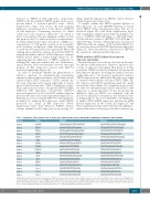
Table 1. Sequences of the primers used to detect gene expression by reverse transcriptase quantitative polymerase chain reaction.
Species
human
human human human human human human human human human human human human human human human human
Name
GAPDH
TRPV1 TRPV4 TRPV5 TRPV6 ALK4 ACVR2 TGFβRI TGFβRII GFRAL ATGL HSL FOXC1 Cav1.3 Cav3.1 Cav3.2 Cav3.3
Forward
CGGAGTCAACGGATTTGGTCGTAT
CAGGCTCTATGATCGCAGGAG CGTCCAAACCTGCGAATGAAGTTC GGTTTTCTACCTAAGGCAGAAGG ACTGACCTCGACTCTCTATGAC GCCATGGGAAGTTGTAATGG GCCACCCTATTACAACATCCTG GAAGAGGACCCTTCATTAG CAACAACATCAACCACAACA ATGGATTCAAAGGGATGTG GCGTGTCAGACGGCGAGAATG CACTACAAACGCAACGAGAC TAAGCCCATGAATCAGCCG CTTCCTCTTCATCATCATCTTC CCACGTGGTCCTTGTCATCA TCGAGGAGGACTTCCACAAG AGGATGAGCTATGACCAGCG
Reverse
AGCCTTCTCCATGGTGGTGAAGAC
TTTGAACTCGTTGTCTGTGAGG CCTCCATCTCTTGTTGTCACTG CTCGAAGCAGTGGAGACTCT GTGGTGATGATAAGTTCCAGCAG GTCCAGGTGCCATTATTCAG GGTCCTGGGTCTTGAGTTG TGCCTCACGGAACCACGAACG TTATAGACCTCAGCAAAGCG TGATAGAAGAACCGTATGGC GCAGCTCGTGGATGTTGGTGG CCAGAGACGATAGCACTTCC GCCGCACAGTCCCATCTCT TCATACATCACCGCATTCC GGGTCAGGAAGATGCGTTCA TGCATCCAGGAATGGTGAG CAGAGAGCAGGGACTCATGC
GAPDH: glyceraldehyde 3-phosphate dehydrogenase; TRPV: transient receptor potential vanilloid; ALK4: activin A receptor type 1B; ACVR2: activin receptor type 2; TGFβRII: trans- forming growth factor-β type II receptor; TGFβRI: transforming growth factor-β type I receptor; GFRAL: GDNF family receptor a-like; ATGL: adipose triglyceride lipase; HSL: hor- mone-sensitive triglyceride lipase; FOXC1: Forkhead box C1; Cav1.3: calcium voltage-gated channel subunit alpha1 C; Cav3.1: calcium voltage-gated channel subunit alpha1 G; Cav3.2: calcium voltage-gated channel subunit alpha1 H; Cav3.1: calcium voltage-gated channel subunit alpha1 I.
haematologica | 2020; 105(11)
2575
