
Page 189 - 2020_11-Haematologica-web
P. 189
Letters to the Editor
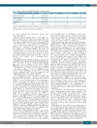
Table 2. SUV in patients with different pathology on biopsy (n=54).
≥5 but <10 10
10
SUV ≥10 max
14
3
Pathology max Richter transformation
Progressive CLL
N Median SUV (range) max
25 11.3 (4.6-24.0)
18 6.4 (1.8-12.5)
SUV max
1
5
<5
SUV max
Second malignancy* 9 8.9 (3.1-17.4) 1 5 3 Inflammation† 2 12.6 (7.4-17.7) 0 1 1
Total 54 8.6 (1.8-24.0) 7 26 21
*Six with recurrent malignancy (two metastatic squamous cell carcinoma of the skin,one each with low grade B-cell lymphoma with plasmacytic differentiation,metastatic melanoma, metastatic Merkel cell carcinoma, and metastatic lung cancer) and three with new malignancy (one each with renal cell carcinoma, soft tissue sarcoma, and metastatic carcinoma of unknown primary origin). †One with reactive gastropathy, the other with acute and chronic granulomatous changes due to herpes simplex virus. SUV: standardized uptake value; CLL: chronic lymphocytic leukemia.
tics between patients who underwent a biopsy versus those who did not.
Fifty-four patients (median SUVmax of 8.6 [range: 1.8- 24.0]) underwent a tissue biopsy. The median time from PET scan to biopsy was 4 days (range: 0-40). The biopsy was targeted towards either the area of maximum SUV (n=29, median SUVmax 7.8) or an alternative area that was easier to access (n=25; median SUVmax 6.7, median SUVmax difference=2.7 compared to the maximum SUV area). The biopsy sites included lymph node (n=34; 10 excisional and 24 core needle), soft tissue mass (n=9; two excisional and seven core needle), bone marrow (n=3), cerebrospinal fluid (n=1) and other organ (n=7; spleen [n=2, splenectomy], kidney, lung, bone [n=1 each, core needle], and stomach [n=2, esophagogastroduo- denoscopy]).
The final pathology was RT in 25 (46%) patients (21 with DLBCL and four with classical Hodgkin lymphoma), CLL in 18 (33%; 15 can be classified as histologically aggressive CLL according to the World Health Organization 2016 criteria, with the presence of expand- ed proliferation centers that are broader than a 20x field or becoming confluent, or a Ki-67 proliferation index >40%), second malignancy in nine (17%; six with recurrent malignancy, three with new malignancy), and inflammation in two (4%) patients (Table 2).
The median SUVmax was 11.3 (range: 4.6-24.0) for patients with RT, 6.4 (range: 1.8-12.5) for patients with progressive CLL (P<0.001 vs. RT; Figure 1A), and 8.9 (range: 3.1-17.4) for patients with a second malignancy (P=0.18 vs. RT; Figure 1A). Only 1 of 7 patients with a SUVmax <5 had RT. In patients with a SUVmax ≥5 but <10, 10 of 26 (38%) had RT; and in patients with a SUVmax ≥10, 14 of 21 (67%) had RT. The sensitivity and specificity for identifying RT (vs. other pathology) using a threshold of SUVmax ≥5 was 96% and 21%, respectively; and using a threshold of SUVmax ≥10 was 56% and 76%, respectively (Figure 1B). The negative predictive value (NPV) of SUVmax <5 in predicting RT was 86%, and the positive predictive value (PPV) of SUVmax ≥10 in predicting RT was 67%. Using the ROC analysis, a threshold of SUVmax ≥9 was determined to be the best discriminator for detecting RT vs. other pathology in all 54 patients who underwent a biopsy, with a sensitivity of 72% and specificity of 72% (Figure 1C). The PPV and NPV of this cut-off was 69% and 75%, respectively. Additional results regarding ibru- tinib hold and survival after PET are available in the Online Supplementary Materials and Methods.
Our study confirms findings by Mato et al.12 In CLL patients receiving a BCRi who underwent a PET scan for evaluation of potential disease progression, although a SUVmax of 9 was the best cut-off to discriminate RT versus other pathology, the sensitivity (72%) and specificity (72%) at this cut-off were both low. The sensitivity
(96%) and NPV (86%) for identification of RT using a lower cut-off of SUVmax ≥5 was excellent, but the speci- ficity (76%) and PPV (67%) remained low using a higher cut-off of SUVmax ≥10. The role of PET in detecting RT needs to be revisited in the novel agent era.
While tissue biopsy still remains the gold standard for diagnosing RT in CLL patients with suspected transfor- mation of disease, a PET scan helps by i) determining if a biopsy should be considered if the SUVmax exceeds a cer- tain cut-off; and ii) identifying the area with the highest FDG uptake for an excisional or core needle biopsy. Mato et al.12 reported a sensitivity of 71% and a specificity of only 4% using a cut-off of SUVmax ≥5 and a sensitivity of 71% and a specificity of 50% using a cut-off of SUVmax ≥10 in CLL patients who progressed after BCRi. In our study, a cut-off of SUVmax ≥5 had a sensitivity of 96% and a specificity of 21%, while a cut-off of SUVmax ≥10 had a sensitivity of 56% and a specificity of 76%. We propose using SUVmax ≥5 as the cut-off to strongly consider biopsy given the high sensitivity (96%) and NPV (86%) in our study. In our cohort, only one patient with SUVmax <5 was diagnosed with RT on excisional biopsy of a cervical lymph node that was enlarging asynchronously. In con- trast, approximately 40% of the patients with a SUVmax ≥5 but <10 and two-thirds of the patients with a SUVmax ≥10 were diagnosed with RT, emphasizing the need to per- form a tissue biopsy in patients with a SUVmax ≥5.
CLL progression on BCRi can be associated with a relatively high SUV in a PET scan, as ibrutinib can change the metabolism of CLL cells by increasing glucose uptake,14,15 and CLL progression developed on ibrutinib is often clinically aggressive.9-12 It is important not to assume a diagnosis of RT even with a high SUVmax (e.g., ≥10), and tissue biopsy is still the gold standard to make a diagnosis.
The strengths of our study include a relatively homo- genous study population from a single institution (CLL patients on BCRi therapy who underwent PET scan for the evaluation of disease progression), and a central review of PET images for SUV measurement/confirma- tion. The limitations include the retrospective design, lack of a tissue biopsy in a subset of patients (although the majority had a low SUVmax), incomplete central pathology review, potential referral bias, and the small cohort size.
In summary, the role of a PET scan in identifying RT in the era of novel agent CLL therapy has evolved owing to the changing biology of CLL with novel targeted therapy. A biopsy should be strongly considered in patients receiv- ing BCRi therapy with suspected RT with a SUVmax ≥5 on PET. Prospective re-examination of the diagnostic value of PET in CLL patients with suspected transformation in the novel agent era with larger cohorts of patients and with central imaging and pathology review is warranted.
haematologica | 2020; 105(11)
2677
