
Page 86 - 2020_09-Haematologica-web
P. 86
A.L. Sørensen et al.
19 had MF (Table 1). One patient with MF developed acute myeloid leukemia and died shortly after inclusion, before initiation of the combination treatment. The patient was excluded from further analysis. The MF patients were in an early stage of disease, with three (17%) having intermediate-2 risk and none having high- risk disease based on the Dynamic International Prognostic Scoring System-Plus (DIPSS+) score. Notably, 47 (94%) of the patients had previously been treated with PEG-IFNa2; 31 (66%) were intolerant, ten (21%) were refractory, and six (13%) were both. Moreover, 27 (54%) had been treated with hydroxyurea, and 25 (50%) had been treated with both hydroxyurea and PEG-IFNa2.
Response evaluations
Of 32 patients with PV, ten (31%) achieved a remission; three (9%) achieved CR, and seven (22%) achieved PR (Figure 1A). Three additional patients achieved BMHR but did not fulfill the criteria for remission; one patient had an increase in total symptom score (TSS); one patient had elevated platelet count, and one patient had both. One patient had progressive disease due to the devel- opment of post-PV MF. Three PV patients dropped out and were classified as having no response. One patient chose not to have a bone marrow biopsy done after 2 years, and five patients had an unsuccessful biopsy. Two of these were in PR.
Of 18 patients with MF, eight (44%) achieved remis- sion; five (28%) achieved CR, and three (17%) achieved PR. Moreover, two (12%) had clinical improvement, both having symptoms response with a ≥50% reduction in TSS (Figure 1A). One additional patient achieved BMHR and PBCR but suffered from grade 1 fatigue and was defined as having stable disease. Two patients had stable disease. Five patients dropped out but did not have pro- gressive disease.
Peripheral blood cell count remission
Of 32 PV patients, five fulfilled the criteria for PBCR at baseline. For the 27 patients who did not have PBCR at baseline, the median time to PBCR was 1 month, and the cumulative incidence of PBCR after 2 years was 0.85 (Figure 1B). Of the five patients with PBCR at baseline, four had PBCR at 2 years. The median duration of the first PBCR was 14 months (Online Supplementary Figure S1), but 12 of the 13 patients who lost PBCR achieved it again during the study period. The proportion of PV patients in PBCR during the study is shown in Figure 1C. Of 14 PV patients in need of phlebotomies within 3 months before inclusion, four needed phlebotomies dur- ing the trial, three of whom required just one phleboto- my. Two additional patients required phlebotomies dur- ing the trial.
Of 18 MF patients, three fulfilled the criteria for PBCR at baseline. For the 15 patients who did not have PBCR at baseline, the median time to PBCR was 3 months, and the cumulative incidence of PBCR after 2 years was 0.73 (Figure 1B). Of the three patients with PBCR at baseline, two had PBCR after 2 years. The median duration until the first PBCR was 5 months (Online Supplementary Figure S1), but seven of the eight patients who lost PBCR achieved it again during the study period. The proportion of MF patients in PBCR during the study is shown in Figure 1D.
Hematocrit, white blood cell count, and platelet count
were all significantly reduced after 2 weeks, and throughout the study period (Figure 1E-G); there were no significant differences between patients with PV and those with MF.
Patient-reported quality-of-life outcomes
During the 2 years of treatment, we observed a statisti- cally significant reduction in MPN-SAF TSS from baseline at all time points except at 1 and 2 years (Figure 2A). The median TSS was reduced from 22 [95% confidence inter- val (95% CI): 16-29] at baseline to 15 (95% CI: 10-22) after 2 years. Compared with patients not achieving remission, patients in remission after 2 years had signifi- cantly (P<0.05) larger reductions in TSS at several time points (figure 2B). The median TSS in patients achieving remission was reduced from 17 (95% CI: 10-27) at base- line to 7 (95% CI: 4-13) after 2 years. The median TSS in patients not achieving remission was 26 (95% CI: 18-37) at baseline and 25 (95% CI: 16-36) after 2 years. The fol- lowing items of the TSS were significantly reduced at more than half of the time points, compared with base- line: early satiety (P<0.05), night sweats (P<0.01), itching (P<0.01) and weight loss (P<0.001) (Figure 2C). We found no significant difference in TSS change between patients with MF and patients with PV.
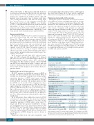
Table 1. Patients’ characteristics at baseline. Polycythemia vera
Myelofibrosis (n=18)
59 (51, 67)
10 (56)
5 (27.8) 6.2 (2.6, 8.0)
6 (33) 9 (50) 3 (17)
-
2 (11) 8 (44) 0 (0)
17 (94) 6 (33) 6 (33) 5 (28)
12 (67)
45 (23, 62)
3 (17)
14.0 (12, 18.5) 24.0 (18.5–41.0) 12.8 (12.0, 13.4) 0.40 (0.35, 0.41) 7.8 (5.4, 10.5) 419 (381, 464)
(n=32)
57 (49, 67)
19 (59)
-
6.9 (2.9, 10.0)
- - -
21 (66) 9 (28) 16 (50) 14 (44)
30 (94) 21 (66) 19 (59) 8 (25)
JAK2 V617F positive, n (%)
JAK2 V617F allele burden (%), median (IQR) 40 (19, 79)
Age (years), median (IQR)
Sex, male (%)
Post-ET or -PV myelofibrosis, n (%) Years since diagnosis, median (IQR)
DIPSS+ score for myelofibrosis patients Low, n (%)
Intermediate-1, n (%)
Intermediate-2, n (%)
High-risk PV, n (%)
Prior thrombosis, n (%)
Constitutional symptoms, n (%)
Need of phlebotomies in last 3 months, n (%)
Prior cytoreductive treatment PEG-IFNa2, n (%)
HU, n (%)
PEG-IFNa2 and HU, n (%) Anagrelide, n (%)
Palpable spleen, n (%)
Spleen size by US (cm), median (IQR) MPN-SAF TSS, median (IQR) Hemoglobin (g/dL), median (IQR) Hematocrit, median (IQR)
WBC count (x 109/L), median (IQR) Platelet count (x 109/L), median (IQR)
6 (19) 14.0 (12.1, 17.3) 21.0 (5.5, 35.0) 13.3 (12.7, 13.9) 0.42 (0.41, 0.44) 8.4 (5.4, 12.7) 401 (251, 592)
32 (100)
IQR: interquartile range; ET: essential thrombocythemia; PV: polycythemia vera; DIPSS+: Dynamic International Prog-nostic Scoring System-Plus; PEG-IFNa2: pegylated interferon-a2; HU, hydroxyurea; US: ultrasonography; MPN-SAF TSS, Myeloproliferative Neoplasm Symptom Assessment Form total symptom score; WBC: white blood cell.
2264
haematologica | 2020; 105(9)
