
Page 53 - 2020_09-Haematologica-web
P. 53
Manifestations of disease in PKD
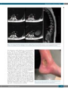
Figure 1. Paravertebral extramedullary hematopoietic masses in pyruvate kinase deficiency. (A) Multiple transverse sections demonstrating paravertebral masses (arrows) in close proximity to nerve roots. (B) Sagittal section demonstrating a large paravertebral mass (bounded by arrows) extending from the vertebra.
liver dysfunction, cardiac dysfunction, or endocrinopathies if not addressed. A small but significant proportion of patients can develop extramedullary hematopoiesis.11 Extramedullary hematopoietic masses in PKD are often par- avertebral,24-26 as illustrated in Figure 1. These masses can enlarge over time and can result in nerve root compression resulting in neurological compromise, including paralysis, if left untreated.27,28 Although evidence is lacking, some mem- bers of the working group treat patients with progressing paravertebral masses with chronic red cell transfusion to suppress the growth of the masses. Extramedullary hematopoietic masses can also be mistaken for malignant tumors, especially in undiagnosed patients. As in other hemolytic anemias, patients with PKD can develop lower- extremity ulcers, usually medial in association with the medial malleolus,29 which can be slow to heal or even fail to heal (Figure 2). There are no data to guide the management of leg ulcers in PKD, so ulcers are managed similarly to those seen in sickle cell disease or thalassemia.30,31
Once PKD has been diagnosed, screening and regular monitoring for complications from chronic hemolysis should be initiated, since many complications, such as iron overload, can be asymptomatic.15 The type and frequency of screening vary between institutions, but the screening is usually directed at complications that carry high morbidity if untreated. These include iron overload, extramedullary hematopoiesis, osteopenia, osteoporosis, gallstones, and pulmonary hypertension. Table 1 shows our consensus approach to screening in patients with PKD.
Figure 2. Lower extremity non-healing ulcer in an adult with pyruvate kinase deficiency. Note the location posterior to the medial malleolus.
haematologica | 2020; 105(9)
2231
