
Page 45 - 2020_09-Haematologica-web
P. 45
Molecular heterogeneity of PK deficiency
PIEZO1 genes, whereas in five other cases, whole genome sequencing identified different intronic variants, all pre- dicted to perturb normal mRNA processing and confirmed by minigene assays.55
Large insertions/deletions
Large indels are rare, possibly due to the technical diffi- culties in identifying them. The most frequent is the dele- tion of the 1149 bp characteristic of the Roma communi- ty, which leads to skipping of exon 11.40 A very large deletion of 5006 bp that results (at the cDNA level) in the loss of exons 4 to 11 (c.283+1914_c.1434del5006) has been described in patients of Vietnamese origin.17,41 A large homozygous insertion of 367 bp (c.939_940ins367) containing an Alu element (AluYb9)57 was identified in two unrelated children with severe transfusion-depen- dent hemolytic anemia, from the Middle East. Other vari- ants have been reported although the exact cut-off point has not been identified (3 of the 5 different large deletions identified within the PKD NHS).35
As for deep intronic variants, the search for large indels may add to the costs of analysis and require techniques not always available; however, it should always be con- sidered when one mutation or no mutations at all are detected in a patient with clinical and biochemical diag- nosis of PK deficiency. The eventuality of a large deletion at the heterozygous level should always be taken into account in patients carrying homozygous pathogenic variants, in whom the allelic transmission has not been confirmed through analysis of the parents.
Inherited pyruvate kinase hyperactivity
Inherited hyperactivity of red blood cell PK (OMIM 102900) has been reported in only three families with apparent asymptomatic conditions with different etiolo- gies.58-60 This rare condition was attributed in the past to a heterozygous mutation in the PKLR gene61 or to the persistent expression of the fetal isozyme PK-M2.59 More recently, there was a report of a family characterized sole- ly by the increased expression of a kinetically normal PK- R, in the absence of mutations in PKLR codifying and reg- ulatory regions as well as variations in PKLR copy num- ber, and exclusion of co-segregation with the PKLR locus;
in this case, the authors postulated that the causative mutation resides in a novel, unidentified locus, responsi- ble for upregulating PKLR gene expression.60
Other genes associated with pyruvate kinase deficiency
KLF1 is a transcription factor involved in terminal ery- throid differentiation, and regulates many of the genes implicated in red cell enzyme deficiencies, including PKLR. Decreased PK activity in the absence of PKLR mutations has been reported in patients who were com- pound heterozygotes for KLF1 variants, possibly leading to misdiagnosis of these cases with PK deficiency;62 these patients displayed severe, transfusion-dependent neona- tal anemia with a broad spectrum of red cell morpholog- ical abnormalities and a remarkable persistence of fetal and embryonic globin synthesis.
Geographical distribution of PKLR variants
PK deficiency has a worldwide geographical distribu- tion. A careful literature review established that the prevalence of clinically-diagnosed PK deficiency was likely between 3.2 and 8.5 cases per million in Western populations, while the prevalence of diagnosed and undi- agnosed PK deficiency could possibly be as high as 51 cases per million.63-65 The causes of this variability can be explained by the rarity of the disorder, the high percent- age of undiagnosed cases and the absence of disease reg- istries and specific population studies.65
There is a high frequency of PK deficiency in Middle East and sub-Saharan Africa populations, possibly due to selective pressure from malaria;39,66 some reports have, in fact, suggested that PK deficiency provides protection against infection and replication of P. falciparum in murine models and in ex vivo experiments with red cells from PK- deficient patients.67-69 Most of the molecular variants reported are private, and it is thus difficult to define a geo- graphical distribution. Despite this, the most frequent mutations in PK deficiency are distributed with a strong ethnic and regional background; in addition, some muta- tions have a high frequency in specific populations as a result of a founder effect (Table 2).
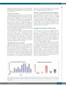
Figure 4. Distribution of variants along the PKLR gene and pyruvate kinase structural domains. Distribution of unique pathogenic variants reported in the Human Genome Mutation Database along exons (left side), and distribution of affected residues in the four different structural domains (right side). aa: amino acid; N: N- terminal domain.
haematologica | 2020; 105(9)
2223
