
Page 30 - 2020_09-Haematologica-web
P. 30
J. Delgado et al.
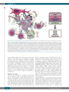
Figure 2. The chronic lymphocytic leukemia microenvironment. Communication between chronic lymphocytic leukemia (CLL) cells and stromal cells, T cells and nurse-like cells (NLC) is established and maintained by direct contacts, chemokine/cytokine receptors, adhesion molecules and ligand-receptor interactions. CLL cells migrate to tissues attracted by the chemokines CXCL12 secreted by NLC and stromal cells, CXCL13 by follicular dendritic cells (FDC), and CCL19/CCL21 by high- endothelial venules, which interact with the CLL receptors CXCR4, CXCR5 and CCR7, respectively. Adhesion molecules (e.g., a4β1 integrin, LFA-1) and their ligands (VCAM1, ICAM, among others) facilitate tumor cell migration and homing. Environmental or auto-/self-antigens and homotypic IG interactions trigger B-cell receptor (BCR) activation capable of driving CLL proliferation.22,138 Interactions between CD40 and CD40 ligand (CD40L) on activated CD4+ T cells are critical in the context of antigen presentation and induction of normal B-cell responses. Activated CLL cells secrete chemokines (CCL2, CCL3, and CCL4) and angiogenic factors that attract T cells and different stromal cells.30,139 Suppressive factors (IL-10)140 and immune inhibitory molecules (PD-L1 among others)141 facilitate tumor cells to evade immune- response and maintain tolerance. Anti-tumor CD8+ T cells become exhausted by constant exposure to tumor-derived antigens leading to cell exhaustion.40 Regulatory T cells (Treg) exert an inhibitory effect on CD4+ and CD8+ cells through secretion of suppressive cytokines.142 Tumor-released extracellular vesicles carrying noncoding RNA and proteins induce an inflammatory phenotype in T cells, monocytes, and stromal cells.143
some banding analysis and chromosomal microarray analysis is the identification of complex karyotypes, observed in up to 20% of patients with CLL. A complex karyotype appears not only prognostic but also predictive in the context of treatment with both conventional and novel agents.54,55 Intriguingly, a subset of patients with complex karyotypes carrying trisomy 12, trisomy 19, and additional trisomies seem to correspond to a particular genetic subgroup with favorable outcome.54–56 In contrast to patients with other hematologic malignancies, patients with five or more aberrations have a worse prognosis compared to those with “less complex” karyotypes (3 or 4 aberrations).55
Mutational landscape
On average, CLL tumors accumulate around 2,500 somatic mutations with a clear difference between M- CLL and U-CLL (3,000 vs. 2,000 somatic mutations on average, respectively). This increased mutation burden observed in M-CLL has limited transformation potential as patients with M-CLL have fewer mutated drivers and better clinical outcomes than patients with U-CLL.3 The mutational landscape of the disease is remarkably hetero- geneous, with only a handful of genes mutated in more
than 5% of patients at diagnosis (NOTCH1, SF3B1, TP53, ATM) followed by a long tail of genes mutated at lower frequencies.3,4 Despite this diversity, most mutated driv- ers cluster in a number of cell pathways (Figure 3), such as NOTCH1 signaling (NOTCH1, FBXW7); BCR and TLR signaling (EGR2, BCOR, MYD88, TLR2, IKZF3); the MAPK-ERK pathway (KRAS, NRAS); NF-κB signaling (BIRC3, NFKB2, NFKBIE, TRAF2, TRAF3); chromatin modifiers (CHD2, SETD2, KMT2D, ASXL1); cell cycle (ATM, TP53, CCND2, CDKN1B, CDKN2A); DNA dam- age response (ATM, TP53, POT1); and RNA splicing and metabolism (SF3B1, U1, XPO1, DDX3X, RPS15). This clustering suggests that mutations belonging to the same cell process may have similar functional and clinical impacts,57,58 but this hypothesis requires further confirma- tion. A detailed descriptions of these cell processes can be found elsewhere.51
This accumulation of low-frequency driver alterations highlights the striking interpatient heterogeneity of CLL, which is partly determined by three major factors: (i) the cell of origin: M-CLL have fewer driver mutations than U- CLL and some mutated genes are almost exclusively or predominantly seen in one of the two subtypes (e.g MYD88 and PAX5 in M-CLL and U1, NOTCH1, and
2208
haematologica | 2020; 105(9)
