
Page 76 - Haematologica Atlas of Hematologic Cytology
P. 76
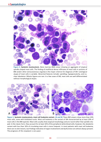
Figure 4. Systemic mastocytosis. Bone marrow (BM) smear showing an aggregate of atypical spindle-shaped mast cells. The finding of multifocal dense infiltrates of mast cells in sections of BM and/or other extracutaneous organ(s) is the ma or criterion for diagnosis of SM. Cytological atypia of mast cells is variable. Abnormal features include: spindling, hypogranularity, and nu- clear lobulation. Mitotic figures are rare. In a few cases of SM, mast cells are well differentiated without morphological atypia.
AB
Figure 5. Systemic mastocytosis  mast cell leu emia variant. (  and  ) These BM smears show more than 20% mast cells, some with bilobated nuclei. Mast cell leukemia is the variant of SM characterized by at least 20% of mast cells in the BM aspirate. Mast cells usually show atypical morphology and are round rather than spindle-sha- ped. In the classic form, they account for at least 10% of the peripheral white blood cells; however, the aleukemic variant with less than 10% of circulating mast cells is more frequent. In most patients with mast cell leukemia, there are no skin lesions, but findings indicative of organ involvement and dysfunction are almost always present. The prognosis of this neoplasm is very poor.
63
