
Page 73 - Haematologica Atlas of Hematologic Cytology
P. 73
CHAPTER 9 - Mastocytosis
Chapter 9. MASTOCYTOSIS
Mastocytosis is characterized by a clonal, neoplastic proliferation of mast cells that accumulate in one or more organs. It includes heterogeneous clinical entities ranging from simple skin lesions that can spontaneously regress to very aggressive forms with involvement of many organs and a severe prognosis. Table 1 reports the classification of the different mastocytosis variants according to the World Health Organization group (Horny et al., 2017).
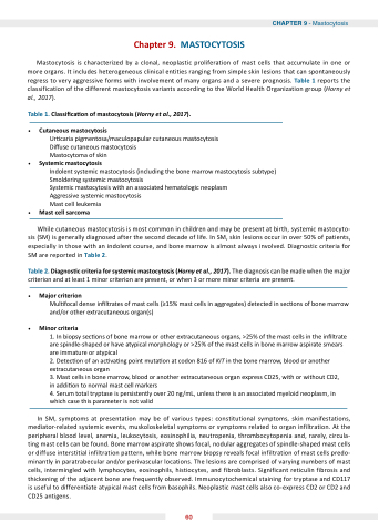
Table 1. lassi ca on of mastocytosis (Horny et al., 2017).
utaneous mastocytosis
r caria pigmentosa/maculopapular cutaneous mastocytosis Di use cutaneous mastocytosis
Mastocytoma of skin
Systemic mastocytosis
Indolent systemic mastocytosis (including the bone marrow mastocytosis subtype) Smoldering systemic mastocytosis
Systemic mastocytosis with an associated hematologic neoplasm
Aggressive systemic mastocytosis
Mast cell leukemia
Mast cell sarcoma
While cutaneous mastocytosis is most common in children and may be present at birth, systemic mastocyto- sis (SM) is generally diagnosed after the second decade of life. In SM, skin lesions occur in over 50% of patients, especially in those with an indolent course, and bone marrow is almost always involved. Diagnostic criteria for SM are reported in Table 2.
Table 2. Diagnos c criteria for systemic mastocytosis (Horny et al., 2017). The diagnosis can be made when the major criterion and at least 1 minor criterion are present, or when 3 or more minor criteria are present.
Major criterion
Mul focal dense in ltrates of mast cells (≥15% mast cells in aggregates) detected in sec ons of bone marrow and/or other extracutaneous organ(s)
Minor criteria
1. In biopsy sec ons of bone marrow or other extracutaneous organs, >25% of the mast cells in the in ltrate are spindle-shaped or have atypical morphology or >25% of the mast cells in bone marrow aspirate smears are immature or atypical
2. Detec on of an ac va ng point muta on at codon 816 of KIT in the bone marrow, blood or another extracutaneous organ
3. Mast cells in bone marrow, blood or another extracutaneous organ express CD25, with or without CD2, in addi on to normal mast cell markers
4. Serum total tryptase is persistently over 20 ng/mL, unless there is an associated myeloid neoplasm, in which case this parameter is not valid
In SM, symptoms at presentation may be of various types: constitutional symptoms, skin manifestations, mediator-related systemic events, muskoloskeletal symptoms or symptoms related to organ infiltration. At the peripheral blood level, anemia, leukocytosis, eosinophilia, neutropenia, thrombocytopenia and, rarely, circula- ting mast cells can be found. Bone marrow aspirate shows focal, nodular aggregates of spindle-shaped mast cells or diffuse interstitial infiltration pattern, while bone marrow biopsy reveals focal infiltration of mast cells predo- minantly in paratrabecular and/or perivascular locations. The lesions are comprised of varying numbers of mast cells, intermingled with lymphocytes, eosinophils, histiocytes, and fibroblasts. Significant reticulin fibrosis and thickening of the ad acent bone are frequently observed. Immunocytochemical staining for tryptase and CD117 is useful to differentiate atypical mast cells from basophils. Neoplastic mast cells also co-express CD2 or CD2 and CD25 antigens.
60
