
Page 247 - Haematologica Atlas of Hematologic Cytology
P. 247
CHAPTER 30 - Hypereosinophilic syndrome
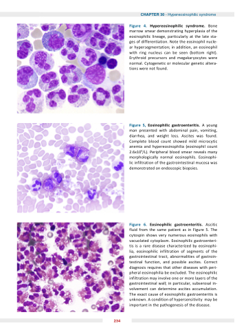
Figure 4 Hypereosinophilic syndrome
Bone marrow smear demonstrating hyperplasia of the eosinophilic lineage particularly at at the late sta- ges of differentiation Note the eosinophil nucle- ar hypersegmentation in in addition an eosinophil with ring nucleus can be seen (bottom right) Erythroid precursors and megakaryocytes were normal Cytogenetic or or molecular genetic genetic altera- tions were not found Figure 5 Eosinophilic gastroenteritis A young man presented with abdominal pain vomiting diarrhea and weight loss Ascites was found Complete blood count showed mild microcytic anemia and hypereosinophilia (eosinophil count 2 6x10⁹/L) Peripheral blood smear reveals many morphologically normal eosinophils Eosinophi- lic infiltration of the gastrointestinal mucosa was demonstrated on on endoscopic biopsies Figure 6 Eosinophilic gastroenteritis Ascitic fluid from the same patient as in Figure 5 The cytospin shows very numerous eosinophils with vacuolated cytoplasm Eosinophilic gastroenteri- tis is is is a a a a a rare disease characterized by eosinophi- lia eosinophilic infiltration of of segments of of the gastrointestinal tract abnormalities of gastroin- testinal function and possible ascites Correct diagnosis requires that other diseases with peri- pheral eosinophilia be excluded The eosinophilic infiltration may involve one or or more layers of the gastrointestinal wall in in in in particular subserosal in- volvement can determine ascites accumulation The exact cause of eosinophilic gastroenteritis is is unknown A condition of hypersensitivity may be important in the the pathogenesis of the the disease 234
