
Page 217 - Haematologica Atlas of Hematologic Cytology
P. 217
CHAPTER 26 - Inherited hemolytic anemias
Chapter 26. INHERITED HEMOLYTIC ANEMIAS
Hereditary hemolytic anemias are a group of heterogeneous diseases mainly caused by abnormalities in red blood cell (RBC) membrane or metabolism. Because of the rarity and heterogeneity of these diseases, diagnosis may be challenging despite the availability of a variety of laboratory tests.
The designation for each of the hereditary RBC membrane disorders denotes the characteristic RBC morpho- logy expected to be found on the patient’s peripheral blood smear. Abnormal RBC morphology is often an indi- cator for, but not necessarily specific to, the RBC disorder under investigation.
Microscopic analysis of the blood film is very useful because it can, in many cases, provide a clue to the dia- gnosis of a particular RBC defect. Moreover, RBC morphology examination can, in a few cases (e.g. RBC membra- ne disorders, sickle cell anemia), provide a definitive diagnosis, although it usually suggests a differential diagno- sis requiring further study. Morphological changes such as basophilic stippling and target cells are not definitively associated with a particular hemoglobinopathy, but may also be suggestive of rare RBC enzymyopathies, such as deficit of pyrimidine 5’nucleotidase deficiency. Thus, despite the advances in automated blood cell counting, the blood film retains a crucial role in the diagnosis of RBC disorders. It should be remembered that RBC morphology examination has the advantage of speed, and this may be important in severe cases of hemolytic anemias that require prompt treatment.
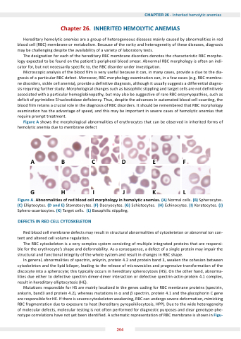
Figure A shows the morphological abnormalities of erythrocytes that can be observed in inherited forms of hemolytic anemia due to membrane defect
ABCDEF GHIJKL
Figure A. Abnormalities of red blood cell morphology in hemolytic anemias. (A) Normal cells. (B) Spherocytes. (C) Elliptocytes. (D and E) Stomatocytes. (F) Dacryocytes. (G) Schistocytes. (H) Echinocytes. (I) Keratocytes. (J) Sphero-acantocytes. (K) Target cells. (L) Basophilic stippling.
DEFECTS IN RED CELL CYTOSKELETON
Red blood cell membrane defects may result in structural abnormalities of cytosketeton or abnormal ion con- tent and altered cell volume regulation.
The RBC cytoskeleton is a very complex system consisting of multiple integrated proteins that are responsi- ble for the erythrocyte’s shape and deformability. As a consequence, a defect of a single protein may impair the structural and functional integrity of the whole system and result in changes in RBC shape.
In general, abnormalities of spectrin, ankyrin, protein 4.2 and protein band 3, weaken the cohesion between cytoskeleton and the lipid bilayer, leading to the release of microvesicles and progressive transformation of the discocyte into a spherocyte; this typically occurs in hereditary spherocytosis (HS). On the other hand, abnorma- lities due either to defective spectrin dimer-dimer interaction or defective spectrin-actin-protein 4.1 complex, result in hereditary elliptocytosis (HE).
Mutations responsible for HS are mainly localized in the genes coding for RBC membrane proteins (spectrin, ankyrin, band3 and protein 4.2), whereas mutations in and spectrin, protein 4.1 and the glycophorin C gene are responsible for HE. If there is severe cytoskeleton weakening, RBC can undergo severe deformation, mimicking RBC fragmentation due to exposure to heat (hereditary pyropoikilocytosis, HPP). Due to the wide heterogeneity of molecular defects, molecular testing is not often performed for diagnostic purposes and clear genotype-phe- notype correlations have not yet been identified. A schematic representation of RBC membrane is shown in Figu-
204
