
Page 58 - 2019_03-Haematologica-web
P. 58
A. Al-Sharea et al.
nolol (Figure 5E). This was paralleled by a decrease in extramedullary hematopoiesis in the spleen as evidenced by fewer proliferating HSPCs, GMPs and less monocytes and neutrophils (Figure 5, F-I). Taken together, these data suggest that lowering responsiveness to chronic sympa- thetic signaling in the BPH/Apoe-/- mice results in an over- all dampening of myelopoiesis.
Blocking sympathetic signalling decreases atherosclerosis in BPH/Apoe–/– mice
To examine if the reduction in sympathetic tone and dampening of myelopoiesis was associated with reduced
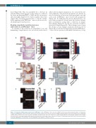
atherosclerotic plaque progression, we assessed the size and complexity of lesions in the proximal aorta. Firstly, we noted a reduction in lesion size in the proximal aorta and aortic arch of BPH/Apoe-/- mice treated with propranolol (Figure 6A, B). Exploring the lesion characteristics, we noted that propranolol treated mice had reduced plaque lipid accumulation along with a reduction in plaque macrophages (Figure 6C, D). We also observed a trend for increased collagen (Figure 6E). These changes were seen in the absence of any changes in plasma cholesterol levels (Figure 6F). Given that the hypertension in the BPH/Apoe- /- mice did not promote endothelial dysfunction, it sug-
AB
CD
EF
Figure 6. Propranolol inhibits plaque progression in BPH/Apoe-/- mice. BPH/Apoe-/- mice were fed a WTD for 16 weeks and treated with vehicle or Propranolol (0.5g/L) in drinking water. At the end point, atherosclerosis in the proximal aorta was assessed for A) H&E staining for plaque size in the proximal aorta and B) lipid content (ORO+) lesions in the aortic arch were quantified. Proximal aortas were also stained for C) lipid content (ORO), D) macrophages (CD68) and E) collagen (picosirius red). Lesions were imaged at X4 (insets) and zoomed in to view single lesion, scale bar = 100 mm. F) Total plasma cholesterol levels. Data are presented as mean ± SEM where *P<0.05 and **P<0.01 (Student’s t-test). A,B) n= 9, C-E) n=7, F) n=9.
464
haematologica | 2019; 104(3)
