
Page 31 - Haematologica Vol. 110 - January 2025
P. 31
REVIEW ARTICLE - Molecular pathogenesis and novel treatments for CMML L. Marando et al.
duction in the diversity of the HSC pool, such that by the age of 70 years, most peripheral blood cells are derived from only 10-20 HSC clones.7 Interestingly, only 20% of the HSC clones remaining in people aged >70 years have identifiable mutations in known driver genes.7 While CHIP is associated with an increased risk of developing hema- tologic malignancies,10-12 the more prevalent, presumed
AB
“driverless” clonal expansions noted in the elderly, might underlie other blood- and immune-related signs of ag- ing.13-15 Notably, while CHIP is present in >90% of individ- uals ≥85 years,8 hematologic malignancies continue to be a rare entity, supporting the hypothesis that extrinsic non-cell-autonomous mechanisms are pivotal in shaping the natural history of CHIP.
CD
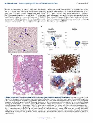
Figure 1. Morphological and immunophenotypic characteristics of chronic myelomonocytic leukemia. Peripheral blood and bone marrow morphology, immunohistochemistry, and cytochemical analysis in chronic myelomonocytic leukemia (CMML). (A) Wright-Gi- emsa ×200 magnification. Peripheral blood smear of a patient with CMML demonstrating promonocytes (bold arrow) along with dysplastic neutrophils (gray arrow). (B) Monocyte repartitioning flow cytometry demonstrating a normal M01 fraction (classical monocytes, CD14+, CD16−) in a healthy control (~80%), while the right scatter plot shows an expanded M01 fraction (>94%) con- sidered characteristic for a diagnosis of CMML. (C) Bone marrow core biopsy of a patient with CMML demonstrating a plasmacy- toid dendritic cell nodule/aggregate (black circle). (D) Image at a higher magnification showing the same nodule brightly positive for CD123 by immunohistochemistry (×1,000 magnification). (E) Cytochemistry on a bone marrow aspirate in a patient with CMML using a dual esterase stain (a-naphthyl butyrate esterase and choloroacetate esterase) demonstrating dysplastic monocytes taking up both colors (blue and brick red). Normal granulocytes stain bright blue, while normal monocytes stain brick red (×400 magnification).
Haematologica | 110 January 2025
23
E
