


Page 205 - Haematologica Vol. 110 - January 2025
P. 205
LETTER TO THE EDITOR B
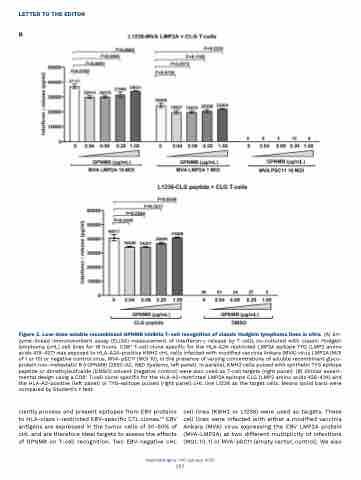
Figure 3. Low-dose soluble recombinant GPNMB inhibits T-cell recognition of classic Hodgkin lymphoma lines in vitro. (A) En- zyme-linked immunosorbent assay (ELISA) measurement of interferon-g release by T cells co-cultured with classic Hodgkin lymphoma (cHL) cell lines for 18 hours. CD8+ T-cell clone specific for the HLA-A24-restricted LMP2A epitope TYG (LMP2 amino acids 419-427) was exposed to HLA-A24-positive KMH2 cHL cells infected with modified vaccinia Ankara (MVA) virus LMP2A (MOI of 1 or 10) or negative control virus, MVA-pSC11 (MOI 10), in the presence of varying concentrations of soluble recombinant glyco- protein non-metastatic B (rGPNMB) (2550-AC, R&D Systems, left panel). In parallel, KMH2 cells pulsed with synthetic TYG epitope peptide or dimethylsulfoxide (DMSO) solvent (negative control) were also used as T-cell targets (right panel). (B) Similar experi- mental design using a CD8+ T-cell clone specific for the HLA-A2-restricted LMP2A epitope CLG (LMP2 amino acids 426-434) and the HLA-A2-positive (left panel) or TYG-epitope pulsed (right panel) cHL line L1236 as the target cells. Means (solid bars) were compared by Student’s t test.
 ciently process and present epitopes from EBV proteins to HLA-class I-restricted EBV-specific CTL clones.1,2 EBV antigens are expressed in the tumor cells of 30-50% of cHL and are therefore ideal targets to assess the effects of GPNMB on T-cell recognition. Two EBV-negative cHL
cell lines (KMH2 or L1236) were used as targets. These cell lines were infected with either a modified vaccinia Ankara (MVA) virus expressing the EBV LMP2A protein (MVA-LMP2A) at two different multiplicity of infections (MOI; 10, 1) or MVA-pSC11 (empty vector, control). We also
Haematologica | 110 January 2025
197


