


Page 202 - Haematologica Vol. 110 - January 2025
P. 202
LETTER TO THE EDITOR AB
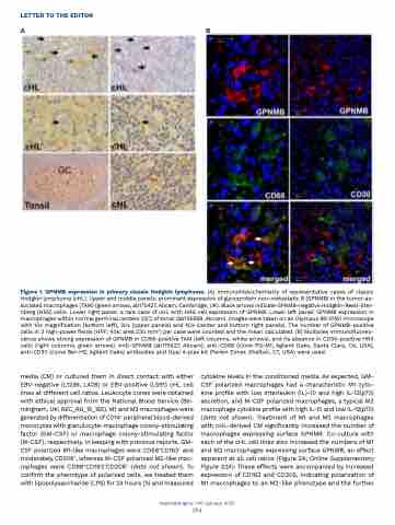
Figure 1. GPNMB expression in primary classic Hodgkin lymphoma. (A) Immunohistochemistry of representative cases of classic Hodgkin lymphoma (cHL). Upper and middle panels: prominent expression of glycoprotein non-metastatic B (GPNMB) in the tumor-as- sociated macrophages (TAM) (green arrows, ab175427, Abcam, Cambridge, UK). Black arrows indicate GPNMB-negative Hodgkin-Reed-Ster- nberg (HRS) cells. Lower right panel: a rare case of cHL with HRS cell expression of GPNMB. Lower left panel: GPNMB expression in macrophages within normal germinal centers (GC) of tonsil (ab125898, Abcam). Images were taken on an Olympus BX-51WI microscope with 10x magnification (bottom left), 20x (upper panels) and 40x (center and bottom right panels). The number of GPNMB-positive cells in 3 high-power fields (HPF; 40x; area 230 mm2) per case were counted and the mean calculated. (B) Multiplex immunofluores- cence shows strong expression of GPNMB in CD68-positive TAM (left columns, white arrows), and its absence in CD30-positive HRS cells (right columns, green arrows). Anti-GPNMB (ab175427, Abcam), anti-CD68 (clone PG-M1; Agilent Dako, Santa Clara, CA, USA), anti-CD30 (clone Ber-H2; Agilent Dako) antibodies and Opal 4-plex kit (Perkin Elmer, Shelton, CT, USA) were used.
 media (CM) or cultured them in direct contact with either EBV-negative (L1236, L428) or EBV-positive (L591) cHL cell lines at different cell ratios. Leukocyte cones were obtained with ethical approval from the National Blood Service (Bir- mingham, UK; REC_RG_15_165). M1 and M2 macrophages were generated by differentiation of CD14+ peripheral blood-derived monocytes with granulocyte-macrophage colony-stimulating factor (GM-CSF) or macrophage colony-stimulating factor (M-CSF), respectively. In keeping with previous reports, GM- CSF polarized M1-like macrophages were CD68+CD163- and moderately CD206+, whereas M-CSF polarized M2-like mac- rophages were CD68+CD163+CD206+ (data not shown). To confirm the phenotype of polarized cells, we treated them with lipopolysaccharide (LPS) for 24 hours (h) and measured
cytokine levels in the conditioned media. As expected, GM- CSF polarized macrophages had a characteristic M1 cyto- kine profile with low interleukin (IL)-10 and high IL-12(p70) secretion, and M-CSF polarized macrophages, a typical M2 macrophage cytokine profile with high IL-10 and low IL-12(p70) (data not shown). Treatment of M1 and M2 macrophages with cHL-derived CM significantly increased the number of macrophages expressing surface GPNMB. Co-culture with each of the cHL cell lines also increased the numbers of M1 and M2 macrophages expressing surface GPNMB, an effect apparent at all cell ratios (Figure 2A; Online Supplementary Figure S2A). These effects were accompanied by increased expression of CD163 and CD206, indicating polarization of M1 macrophages to an M2-like phenotype and the further
Haematologica | 110 January 2025
194


