
Page 71 - Haematologica-5
P. 71
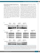
C
Figures 2. DCC-2618 inhibits phosphorylation of KIT and other targets in neoplastic mast cells. (A,C) HMC-1 and ROSA cells were incubated in control medium (ROSAKIT WT: Iscove modified Dulbecco medium (IMDM) with stem cell factor, SCF; ROSAKIT D816V: IMDM without SCF) or medium containing various concentrations of DCC-2618, as indicated, at 37°C for 4 h. Thereafter, cells were harvested and Western blotting was performed as described in the text using antibodies against phosphorylated (p)KIT, total KIT, pBTK and total BTK. (B) HMC-1 and ROSA cells were first pre-incubated overnight in IMDM devoid of fetal calf serum and of SCF. Cells were then treated with DCC-2618 (0.001-10 μM) for 90 min at 37°C. At the end of the treatment, ROSAKIT WT cells were stimulated with SCF (10% CHO-KL) at room temperature for 10 min. Thereafter, cells were harvested and Western blotting was performed as described in the text using antibodies against pSTAT5, total STAT5, pAKT, total AKT, pERK1/2, total ERK1/2. Western blot experiments were performed at least twice. Western blots in this figure show one representative experiment.
DCC-2618: a new drug against mastocytosis
DCC-2618 inhibits IgE-dependent histamine release from basophils and spontaneous tryptase release from neoplastic mast cells
Since patients with SM often suffer from symptoms caused by mediators released from neoplastic MC and/or basophils, we evaluated the effect of DCC-2618 on anti- IgE-induced histamine release. We found that DCC-2618 (0.1-1.0 μM) slightly inhibited anti-IgE mediated histamine release from normal human blood basophils (Figure 4A). This drug effect was found to be specific in that DCC-2618 did not inhibit C5a- or calcium ionophore-induced hista- mine release from basophils (Online Supplementary Figure S4A). As expected, DCC-2618 did not affect the viability of basophils between 0.1 and 1.0 μM and did not induce his- tamine secretion within 30 min of incubation (Online Supplementary Figure S4B). In consecutive experiments, we also found that DCC-2618 suppresses the spontaneous (baseline) secretion of tryptase from HMC-1.1 and HMC- 1.2 cells during the entire incubation period (days 1 through 6) (Figure 4B).
A
B
DCC-2618 counteracts growth and survival of leukemic monocytes and blast cells
We next explored the effects of DCC-2618 on AHN cell- types. In a first step, we examined AML cell responses. DCC-2618 was found to inhibit the proliferation of all AML cell lines tested, with considerably lower IC50 values obtained with the FLT3-mutated cell lines MOLM-13 (132±95 nM) and MV4-11 (147±88 nM) compared to KG- 1 and U937 cells (Table 2, Figure 5A). Similar effects were seen with DP-5439 (Figure 5A). DCC-2618 was also found to induce apoptosis in MOLM-13, MV4-11 and KG-1 cells (Figure 5B and Online Supplementary Figure S5). Finally, we found that DCC-2618 and DP-5439 produced dose-depen- dent inhibition of growth in primary leukemia cells obtained from patients with AML or CMML (Online Supplementary Table S1 and Figure 5C). In one patient with ASM-CMML, we isolated mononuclear cells and found that DCC-2618 and DP-5439 induced growth inhibition of these cells in the same way as in mononuclear cells obtained from patients with CMML without SM (Table 1
haematologica | 2018; 103(5)
803
