
Page 47 - Haematologica-5
P. 47
Stem cell mobilization in sickle cell patients
Introduction
Sickle cell disease (SCD) is caused by a point mutation in the coding region of the HBB (β-globin) gene. As a result, an abnormal β-globin protein is incorporated into hemoglobin tetramers. These mutant tetramers polymer- ize when the local oxygen tension is low. The sickle hemoglobin (HbS) polymers rigidify red blood cells, change these cells’ shape, and are responsible for structur- al damage to the red blood cell membrane. In turn, this modifies the cells’ rheological properties, alters their flow in the microcirculation, and thus causes ischemia, stroke, multi-organ damage, severe acute and chronic pain, and chronic hemolytic anemia. Progressive chronic organ com- plications become the main cause of morbidity and mor- tality in the third decade of life.1 SCD is endemic in Africa, and the World’s Health Organization considers that 7% of the world population carries the trait.
The only curative treatment for SCD is allogeneic hematopoietic stem cell transplantation (HSCT) from matched sibling donors; the disease-free survival rate 6 years after transplantation is reportedly >90%.2,3 Given the limited availability of suitable donors and the increase in toxicity with age, HSCT is only applied with great cau- tion in adult SCD patients (the main target population for curative treatment).
We recently demonstrated that gene therapy is applica- ble to SCD patients, and that the associated toxicity and morbidity rates seem to be lower than those for allogeneic HSCT, at least in the first treated patient.4
As is the case with all genetic diseases, the success of gene therapy in SCD relies on several key factors; these include the source, quality and number of transduced cells, the choice of the conditioning regimen, the level of therapeutic transgene expression, and the quality of the bone marrow (BM) microenvironment at the time of har- vest and transplantation. It is generally acknowledged that 2 to 3x10⁶ CD34+ hematopoietic stem and progenitor cells (HSPC)/kg are required for a successful outcome in autol- ogous HSCT.5 Considering the typical proportion of HSPC that can be corrected in gene therapy clinical trials (~50% of CD34+ HSPC) and an average recovery of 70% of CD34+ cells post-selection, a minimum harvest of ~6x10⁶ CD34+ cells/kg would be required. For reasons that have not been completely elucidated, as for thalassemic
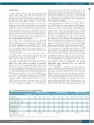
Table 1. Clinical parameters of patients treated with Plerixafor.
SCD Pler 1 (19 years)
patients,6-7 the recovery of HSPC from SCD patients’ BM is peculiarly low (M. Cavazzana, unpublished data). In our ongoing gene therapy trial (HGB-205, ClinicalTrials.gov number, NCT02151526), two BM harvests (each requiring an exchange transfusion program before general anesthe- sia) were needed to obtain enough cells to transplant the three SCD patients enrolled.4
Mobilization with granulocyte colony-stimulating fac- tor (G-CSF, Filgrastim) is widely used to increase the har- vest of HSPC (relative to that obtained in a BM aspirate). However, attempts to mobilize HSPC with Filgrastim in SCD have led to severe adverse events, which hamper the cytokine’s use in this setting. The first report of a severe adverse event following mobilization with low-dose Filgrastim (2.5 μg/kg/day) concerned a SCD patient who developed acute chest syndrome and an elevated white blood cell count (63,400/mm3) as early as 3 days after the first injection.8 The temporal relationship between Filgrastim administration, the rapid rise in the white blood cell count and the severe adverse event were strongly sug- gestive of a causal link. Two other severe adverse events (multi-organ failure and a death) were reported in 2001.9-10 Between 2003 and 2008, eight other patients requiring autologous HSCT for a malignant hematopoietic disease were mobilized with Filgrastim after additional precau- tions had been taken: reduction of the HbS level to below 30% via red blood cell exchange, and carefully monitoring of peripheral leukocytosis and the blood ion profile.11-13 Although all eight patients experienced bone pain, hyper- tension or migraine, no severe adverse events were report- ed - providing evidence that SCD patients can be mobi- lized without any major complications. The median [interquartile range] circulating CD34+ cell count was 24.4/μL [21.2-48.6].12-14
As an alternative to Filgrastim, Plerixafor (formerly known as AMD3100) can effectively mobilize HSPC; the CD34+ cell count in the circulation can reach 15 to 40/μL, depending on the dose and the study.15,16 In contrast to Filgrastim (which acts by activating monocytes and neu- trophils in both peripheral blood and the BM), Plerixafor directly inhibits the binding of stroma-cell-derived factor- 1a to its CXC chemokine receptor on HSPC – thus releas- ing stem cells from the BM niches. This mechanistic dif- ference explains the small increase in the white blood cell count and the short time interval between Plerixafor
SCD Pler 2 (20 years) SCD Pler 3 (21 years) Normal values D-1 D+1 D+7 D+30 D-1 D+1 D+7 D+30 D-1 D+1 D+7 D+30
HbS (HPLC G8) 0% 12·1% 12·6% 15·5% 36·0% 20·2% 15·8% ND 37·1% 6·2%*,** 13·2%** 25·2%** 36·8%**
Total bilirubin (mol/L) 0-17 112 105 109 132 32 35 ND Conjugatedbilirubin(mol/L) 0-5 8 7 8 9 11 13 ND
32 30 30 31 53 12 10 10 10 6
Lactate dehydrogenase (U/L) White blood cells (109/L) Neutrophils (109/L) Monocytes (109/L)
125-243 4·0-10·0 1·5-7 0·2-1 22-275 < 36
399 409 361 429 9·9 20·1 11·8 13·2 4·9 14·9 7·8 8·2 2·0 2·4 1·7 2·3 2441 ND 2674 3111
340 (±50)
566 584 ND 11·9 11·2 ND 7·6 9·2 ND 0·9* 0·9 ND
ND 1009 1473 120 (±30)
605 6·2 2·6 0·7 1962
378 383 426 501 14·0 11·3 9·6 10·4 9·6 9·3 6·4 6·5 1·8* 1·3 1·5 1·8 ND 381 365 446
55 (±30)
Ferritin (μg/L)
Liver quantification
(at inclusion period) (μmol/g)
HPLC: High-performance-liquid-chromatography; D: day. ND: not determined. * D+3. **Capillary 3 (and not G8).
haematologica | 2018; 103(5)
779
