
Page 138 - Haematologica-5
P. 138
A. Agathangelidis et al.
A
B
C
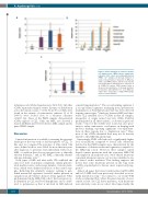
Figure 3. Exonic mutations in our monoclonal B- cell lymphocytosis (MBL)/chronic lymphocytic leukemia (CLL) cohort and polymorphonuclear (PMN) cell samples. (A) Average numbers of exonic non-synonymous mutations in MBL/CLL entities and PMN samples. (B) The vast majority of non-synonymous mutations were missense in all 3 MBL/CLL entities. PMN samples failed to show such predominance. (C) Average numbers of mutations with VAF≥50% for all 3 MBL/CLL entities were comparable. A single PMN sample carried a clonal mutation.
870
technique each (Online Supplementary Table S16). All other sCNAs represented unique events. In terms of distribution across the chromosomes, 7 of the 32 (21.9%) sCNAs were found in the vicinity of centromeres, whereas 11 of 32 (34.4%) were located close to a telomere (distance <10x107 bp). None of the PMN samples demonstrated sCNAs typical of CLL. Only one MBL case showed a shared del(8)(p11.22) between the LC-MBL sample and its paired PMN sample.
Discussion
Limited information is available concerning the genomic landscape at the very early or indolent phases of CLL. To this end, we compared the genomes of ultra-stable CLL cases, defined as those cases stable for more than ten years after diagnosis, to genomes from individuals with: i) LC- MBL, a condition that does not progress into a clinically relevant leukemia;26 and, ii) HC-MBL, a clinically identifi- able pre-leukemic state.25
Both types of MBL and ultra-stable CLL exhibited the same low level of genomic complexity, similar genome- wide mutation rates, and average number of exonic muta- tions, which were distinct from those of the control sam- ples. Reflecting this similarity, analysis relating to pub- lished mutational signatures revealed similar patterns in samples from all 3 entities. In more detail, signature 9 that predominated in the MBL/CLL cohort has been previous- ly identified in CLL and B-cell lymphomas and is attrib- uted to polymerase η that is involved in AID-induced
somatic hypermutation.16 The second ranking signature 1 is an age-related signature stemming from spontaneous deamination of 5-methylcytosine that has been detected in many cancer types.16 Analogies between MBL and ultra- stable CLL extended also to sCNAs in that all samples, irrespective of origin, carried very few sCNA. Del(13q) predominated in all three entities, as shown in previous studies.26 Most of the sCNAs were located in close prox- imity to either centromeres or telomeres, in keeping with previous findings reporting significant over-representa- tions in these regions due to duplication rates.43 Thus, most of the sCNAs identified here may not be directly related to the MBL/CLL phenotype.
Interestingly, PMN cells harbored a significantly higher load of mutations compared to buccal cells. Mutations detected in the PMN samples were characterized by the dominance of distinct mutational signatures compared to the MBL/CLL cohort. However, these samples carried shared somatic mutations with the respective MBL/CLL cell samples in all analyzed cases. Most shared mutations concerned intergenic regions, yet we also identified a sin- gle shared exonic mutation. This finding supports the notion that some mutations present in the CLL clone could be acquired prior to disease onset, as previously sug- gested.32
Almost all genes that were found mutated in HC-MBL and/or LC-MBL had been previously described as recur- rently mutated in CLL.9,37 In contrast to our recent WES study on relapsing CLL,8 where the great majority of cases carried at least one CLL driver mutation, such mutations were relatively scarce in our cohort. Most importantly, the
haematologica | 2018; 103(5)
